Prepare for University Studies & Career Advancement
Bio-Optics
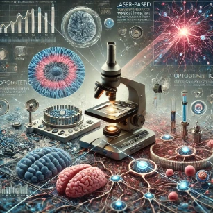
Bio-optics is the study of how light interacts with living systems. It connects Physics, biology, medicine, engineering, and data analysis by asking a powerful question: what can light reveal about cells, tissues, organs, biomolecules, and living processes?
This page belongs to the wider Light and Optics cluster. It shows how ideas such as absorption, scattering, fluorescence, reflection, refraction, interference, polarisation, and photon energy become practical tools for biological observation, medical diagnostics, microscopy, tissue imaging, biosensing, and therapy.
Bio-optics is not simply “using light in biology.” It is a way of turning light into biological evidence. A fluorescent molecule can reveal where a protein is located. A microscope can show cells that ordinary vision cannot resolve. Optical coherence tomography can create layered tissue images. A pulse oximeter can estimate oxygen-related information from the way blood absorbs red and infrared light.
Bio-optics also overlaps with the broader field of Medical Imaging, but the focus here is more specific. Medical Imaging covers many clinical technologies, including X-ray, CT, MRI, ultrasound, nuclear imaging, endoscopy, and optical systems. Bio-optics focuses on how light itself interacts with biological matter and becomes a tool for seeing, measuring, detecting, and sometimes treating living systems.
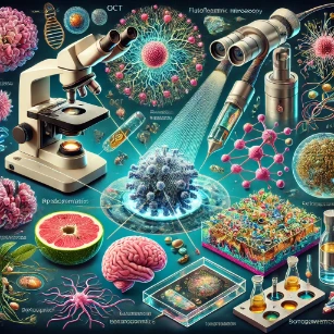
Bio-optics connects light with living systems, showing how optical tools can reveal cells, tissues, biological signals, diagnostic markers, and medical information that ordinary vision cannot see.
What Bio-Optics Really Is
Bio-optics studies the interaction between light and biological matter. This includes cells, tissues, blood, skin, eyes, nerves, plants, microorganisms, proteins, DNA, fluorescent labels, and medical samples. The field asks how light is absorbed, scattered, reflected, transmitted, emitted, delayed, or changed by living materials.
The same optical interaction can have different meanings depending on context. Absorption may reveal blood oxygenation. Scattering may reveal tissue structure. Fluorescence may reveal a labelled protein. Interference may reveal tissue layers. A spectrum may reveal a molecular fingerprint. Bio-optics therefore sits between physical theory and biological meaning. Physics explains what light does. Biology explains what the signal means.
Learning Pathway: Bio-Optics Cluster
This hub organises Bio-Optics into four focused subpages. The pathway begins with molecular and cellular imaging, moves into tissue-layer imaging, then broadens into microscopy and medical diagnostics. Use the links below to navigate the cluster module:
See how absorption, scattering, fluorescence, spectroscopy, OCT, endoscopy, and optical sensors support diagnostic measurement.
Key Concepts in Bio-Optics
Bio-optics is built on several recurring ideas. These ideas appear across microscopy, OCT, fluorescence imaging, spectroscopy, biosensing, medical diagnostics, and therapy.
Light-Tissue Interaction
When light enters biological tissue, it does not simply pass through like it would through empty space. Tissue contains water, proteins, membranes, blood, pigments, fibres, cells, organelles, and microscopic boundaries. These structures alter the path, intensity, colour, phase, and polarisation of light.
Interaction
What Happens
Why It Matters in Bio-Optics
Absorption
Molecules take up light energy
Useful for measuring pigments, haemoglobin, water, oxygenation, and molecular concentration
Scattering
Light is redirected by tissue structures
Affects imaging depth, contrast, tissue appearance, and optical diagnostics
Fluorescence
A molecule absorbs light and emits longer-wavelength light
Useful for labelling cells, proteins, organelles, biomarkers, and biological processes
Reflection
Light returns from a surface or internal boundary
Useful in endoscopy, surface inspection, tissue imaging, and OCT
Refraction
Light bends when entering a medium with a different refractive index
Important in lenses, the eye, optical fibres, tissue interfaces, and focusing systems
Interference
Light waves combine depending on phase and path difference
Central to optical coherence tomography and other precision optical methods
Wavelength Selection and the Optical Window
Wavelength choice is central to bio-optics. Shorter wavelengths can give better resolution but may scatter more strongly or carry higher photon energy. Longer wavelengths, especially in the red and near-infrared range, may penetrate deeper into some tissues but often provide lower resolution. The best wavelength depends on the specimen, the depth needed, the contrast mechanism, the detector, and safety limits.
Optical Contrast, Resolution, and Depth
Biological structures are often transparent or visually similar under ordinary light. Bio-optics therefore depends heavily on contrast. Contrast may come from natural colour, absorption differences, scattering differences, fluorescent labels, phase shifts, polarisation changes, or computational processing.
Bio-optical systems often face a trade-off between resolution and depth. Microscopy can reveal fine cellular detail but usually works best with thin or accessible samples. Diffuse optical methods and near-infrared sensing can probe deeper but usually with less spatial detail. This trade-off is one of the most important design problems in biological optical imaging.
Theoretical Framework: The Beer-Lambert Law, Turbid Media Transport, and the Abbe Resolution Limit
To understand how light propagates through living networks with academic rigor, students must grasp the mathematical rules governing attenuation, turbid scatter, and wave optics limitations.
The Beer-Lambert Law of Absorption
When a beam of light passes through a non-scattering, absorbing biological solution, its intensity decreases exponentially. The change in intensity is described by the linear absorption coefficient μa, which is proportional to the concentration C of the absorbing molecule and its molar absorptivity ε:
I(z) = I0 e−μa z
Where I0 is the initial intensity entering the sample, z is the path distance traveled through the tissue, and μa = ε C. This relationship allows diagnostic tools like pulse oximeters to calculate chemical metrics—such as the ratio of oxygenated to deoxygenated hemoglobin—by monitoring absorption changes at two different wavelengths.
Light Transport in Turbid Media
Most biological tissues are not perfectly clear; they are turbid media where scattering dominates. To track light behavior in these environments, scientists must combine the absorption coefficient with a linear scattering coefficient μs and an asymmetry parameter g. The parameter g measures the average cosine of the scattering angle, representing how forward-directed the scattering is. These values are combined into the reduced scattering coefficient μs′:
μs′ = μs(1 − g)
When scattering is much stronger than absorption (μs′ » μa), the transport of light can be modeled as a diffusion process. The penetration depth of this diffuse light is governed by the effective attenuation coefficient μeff:
μeff = √(3 μa (μa + μs′))
In the near-infrared spectrum between **650 nm** and **950 nm**—often called the optical window of tissue—both absorption and scattering are minimized. This allows light to penetrate centimeters into tissue, enabling deep-layer brain and muscle oxygenation measurements.
The Abbe Diffraction Limit of Resolution
While near-infrared light allows deep tissue penetration, wave optics places a fundamental constraint on spatial resolution. Due to the diffraction of light waves passing through a finite circular lens aperture, a point source forms an Airy disk pattern rather than a perfect point. The minimum distance d required to resolve two distinct structures under a microscope is limited by the wavelength λ and the numerical aperture NA of the objective lens:
d = 0.61 λ / NA
Where NA = n sin(θ), with n tracking the refractive index of the medium, and θ matching the semi-angle of the light collection cone. This diffraction barrier demonstrates why conventional optical microscopes cannot resolve details smaller than roughly half the wavelength of the light used.
Optical Imaging Techniques in Bio-Optics
Bio-optics includes many imaging methods. Some reveal cells and tissues at microscopic scales. Others image tissue layers, blood flow, surface features, or molecular signals.
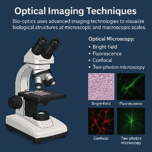
Optical Microscopy: Uses lenses, illumination, contrast methods, and sometimes fluorescent labels to observe cells, tissues, microorganisms, and biological structures. Brightfield, darkfield, phase contrast, fluorescence, confocal, multiphoton, and super-resolution methods all provide different ways of seeing biological samples.
Optical microscopy uses different contrast and imaging methods to reveal biological structures, from ordinary brightfield views to fluorescence, confocal, and multiphoton imaging.
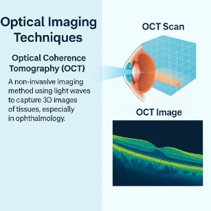
Optical Coherence Tomography: Uses low-coherence light and interference to produce cross-sectional images of biological tissue. It is especially important in ophthalmology because the eye is optically accessible and contains layered structures such as the retina and cornea.
Optical coherence tomography uses reflected light and interference to build cross-sectional images of tissue layers, especially in eye imaging and biomedical research.
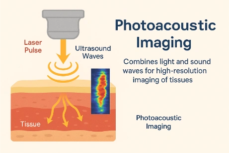
Photoacoustic Imaging: Combines light and sound. A short light pulse is absorbed by tissue, causing a tiny local expansion that generates ultrasound waves. These sound waves can be detected and used to reconstruct images related to optical absorption inside tissue. This method is important because it connects optical contrast with ultrasound detection, providing clear images of blood vessel structure and oxygenation.
Photoacoustic imaging uses light absorption to generate ultrasound signals, combining optical contrast with acoustic detection for tissue imaging.
Endoscopy and Fibre-Optic Imaging: Endoscopy uses optical systems to view internal body surfaces. Optical fibres, miniature cameras, lenses, and illumination systems allow light to reach regions that cannot be seen directly from outside the body. This application connects Bio-Optics with fiber devices, guiding light through narrow transparent pathways for real-time internal diagnosis.
Spectroscopy in Bio-Optics
Spectroscopy studies how light changes with wavelength after interacting with biological matter. Instead of forming only an image, spectroscopy produces a spectrum. That spectrum can reveal chemical composition, molecular structure, oxygenation, fluorescence, or tissue condition.
Spectroscopic Method
Basic Idea
Bio-Optical Use
Absorption spectroscopy
Measures wavelengths absorbed by molecules
Useful for haemoglobin, pigments, oxygenation, and concentration measurements
Fluorescence spectroscopy
Measures emitted light after excitation
Useful for fluorescent probes, biomarkers, and cellular processes
Raman spectroscopy
Measures small wavelength shifts from molecular vibrations
Useful for chemical fingerprinting of tissues, cells, and samples
Near-infrared spectroscopy
Measures interaction with near-infrared light
Useful for tissue oxygenation and selected monitoring applications
Diffuse reflectance spectroscopy
Measures light reflected after scattering through tissue
Useful for tissue composition, blood content, and structural changes
Therapeutic Applications and Optogenetics
Bio-optics is not limited to imaging and diagnosis. Light can also be used therapeutically when wavelength, power, duration, and targeting are carefully controlled.
Photodynamic Therapy (PDT): Uses a light-sensitive substance that is activated by light. When activated in the presence of oxygen, it can produce reactive species that target and destroy diseased cells, such as in localized cancer treatments.
Laser Surgery: Lasers deliver precise, concentrated light for cutting, coagulating, reshaping, or removing tissue. Their usefulness depends on wavelength, power, focusing, pulse duration, tissue absorption, and safety control. This precision allows surgeons to operate on delicate tissues, like the retina, with minimal damage to surrounding areas.
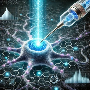
Optogenetics: Uses light-sensitive proteins to control selected cells with light. It is especially important in neuroscience, where researchers can activate or inhibit specific groups of neurons in experimental systems. Optogenetics shows how bio-optics can move beyond observation. Light can become a tool not only for seeing biological activity, but also for controlling it in carefully designed experiments.
Optogenetics uses light-sensitive proteins to control selected cells, making it a powerful example of how optical tools can influence biological activity.
Applications of Bio-Optics
Bio-optics is widely used because light can be sensitive, precise, fast, and compatible with many biological systems. Its applications range from basic research to diagnostics, therapy, environmental monitoring, and biomedical engineering.
Medical Imaging
Bio-optical methods such as OCT, fluorescence imaging, endoscopy, and photoacoustic imaging contribute to the wider field of medical imaging.
Microscopy and Cell Biology
Light microscopy, fluorescence, confocal imaging, and super-resolution methods reveal cells, organelles, proteins, and biological processes.
Medical Diagnostics
Optical sensing, spectroscopy, fluorescence assays, OCT, and pulse oximetry can support diagnostic measurement and monitoring.
Cancer Research and Treatment
Fluorescence imaging, optical biopsies, photodynamic therapy, and image-guided procedures can support cancer research and selected treatments.
Neuroscience
Calcium imaging, fluorescence microscopy, and optogenetics help researchers study and control neural activity in experimental systems.
Optical imaging can monitor cells, scaffolds, tissue growth, structure, and regeneration without always destroying the sample.
Wearable and Point-of-Care Sensors
Optical sensors can measure signals such as blood oxygenation, pulse patterns, fluorescence, colour change, or biomarker response.
Environmental Biosensing
Bio-optical sensors can help detect pathogens, toxins, algae, pollutants, or biological changes in water, plants, and environmental samples.
Bio-optics supports medical imaging, diagnostics, cancer research, neuroscience, tissue engineering, biosensing, and environmental monitoring through the careful use of light.
Connections with Wider Physics and Technology
Bio-optics draws from several parts of physics and technology. These connections help students see why optical biology is not an isolated topic, but a practical meeting point of many fields within the broader framework of Light and Optics.
Visible light, ultraviolet light, and infrared radiation are electromagnetic waves. Their wavelength, frequency, and energy determine how they interact with biological matter.
Diffraction, interference, coherence, resolution, and phase contrast explain the absolute limits and unique strengths of advanced cellular imaging methods.
Lasers provide highly controlled light channels essential for fluorescence excitation, OCT configurations, Raman spectroscopy diagnostics, precision surgery, and targeted optogenetics.
Photonics systems provide the active light sources, high-sensitivity photodetectors, optical filters, cameras, and integrated micro-optical components used to capture biological metrics.
The eye itself functions as a complex biological optical framework, directly connecting core bio-optics with vision science, corrective lens calculations, and high-resolution retinal mapping.
Environmental bio-optics connects cellular plant reflectance, aquatic water quality signatures, photosynthetic tracking, and wide-area ecosystem sensing with quantitative optical monitoring models.
Why Study Bio-Optics?
It Reveals Hidden Biological Structure: Microscopy, fluorescence, OCT, and spectroscopy allow students to see cells, tissues, proteins, blood vessels, and tissue layers that ordinary vision cannot reveal.
It Connects Physics with Medicine: Photon energy, wavelength, scattering, absorption, and interference become practical tools in diagnostics, monitoring, imaging, and treatment.
It Supports Early Detection: Optical techniques can detect subtle structural, chemical, or molecular changes that may be useful in research and medical diagnostics.
It Builds Imaging Literacy: Students learn that images are not neutral pictures. They are shaped by optics, detectors, contrast methods, software, and interpretation.
It Encourages Interdisciplinary Thinking: Bio-optics brings together physics, biology, chemistry, medicine, engineering, computer vision, and data science.
It Opens Career Pathways: Bio-optics supports careers in biomedical engineering, microscopy, medical device design, labs, and clinical technology development.
Frequently Asked Questions: Bio-Optics
What is bio-optics?
Bio-optics is the study of how light interacts with living systems, including molecules, cells, tissues, organs, plants, microorganisms, and medical samples. It combines optics, biology, medicine, engineering, and data analysis to support imaging, diagnostics, therapy, sensing, and biological research.
How does light interact with biological tissues?
Light can be absorbed, scattered, reflected, refracted, transmitted, or emitted as fluorescence. Tissue composition, water content, blood, pigments, fibres, and cell structure all influence how light behaves. These interactions determine imaging depth, contrast, diagnostic signal, and safety.
What are the main imaging methods in bio-optics?
Important methods include optical microscopy, fluorescence imaging, confocal microscopy, multiphoton microscopy, optical coherence tomography, photoacoustic imaging, endoscopy, and spectroscopic imaging. Each method uses light differently to reveal biological structure or function.
What is the difference between fluorescence and bioluminescence?
Fluorescence occurs when a molecule absorbs external light and then emits longer-wavelength light. Bioluminescence is light produced by a chemical reaction inside a living organism, such as in fireflies or some marine organisms. Both can serve as biological optical signals.
What is optical coherence tomography?
Optical coherence tomography, or OCT, uses low-coherence light and interference to create cross-sectional images of tissue. It is especially important in eye imaging because it can show retinal and corneal layers without physically cutting the tissue.
How is bio-optics used in eye and vision science?
Bio-optics supports vision science through retinal imaging, OCT, wavefront measurement, fundus imaging, slit-lamp examination, visual optics, and studies of how the eye focuses and detects light. The eye is one of the clearest examples of a biological optical system.
How does bio-optics contribute to neuroscience?
Bio-optics contributes to neuroscience through fluorescence imaging, calcium imaging, optogenetics, fibre-optic probes, and optical methods for observing or controlling neural activity in research settings. These tools help scientists study neural circuits and brain function.
What are optical biosensors?
Optical biosensors detect biological molecules or processes by measuring changes in light intensity, colour, wavelength, phase, fluorescence, or refractive index. They can be used in medical diagnostics, environmental monitoring, biotechnology, and point-of-care testing.
How is light used therapeutically in medicine?
Light can be used in laser surgery, photodynamic therapy, selected dermatological procedures, ophthalmic treatment, and light-activated biological methods. Therapeutic use requires careful control of wavelength, intensity, exposure time, targeting, and safety.
Are there safety concerns in bio-optics?
Yes. Lasers, ultraviolet light, intense illumination, and some optical procedures can affect the eyes, skin, or living tissue. Bio-optical work must follow suitable safety standards, exposure limits, protective procedures, and ethical guidelines.
What background knowledge is useful for studying bio-optics?
Useful background includes light and optics, electromagnetic waves, wave optics, lenses, photon energy, basic biology, cell structure, physiology, biochemistry, instrumentation, statistics, and image analysis. Programming and data skills are increasingly important.
What careers are linked to bio-optics?
Bio-optics connects to biomedical engineering, medical device design, microscopy, photonics, ophthalmic technology, biotechnology, neuroscience, diagnostics, tissue imaging, optical instrumentation, laboratory research, and clinical technology development.
Review Questions and Answers
Review Questions: Bio-Optical Systems
1. What is bio-optics?
Bio-optics is the study of how light interacts with biological materials and living systems, including absorption, scattering, fluorescence, reflection, imaging, sensing, diagnostics, and therapy.
2. How does light interact with biological tissue?
Light can be absorbed, scattered, reflected, refracted, transmitted, or emitted. These interactions depend on tissue composition and structure, and they determine imaging contrast, penetration depth, and diagnostic signal.
3. What role does fluorescence play in bio-optical imaging?
Fluorescence allows selected molecules, cells, organelles, or markers to emit light after excitation. This creates strong contrast and helps reveal specific biological structures or processes.
4. How is OCT used in medical diagnostics?
OCT uses low-coherence light and interference to create cross-sectional tissue images. It is widely used in eye imaging to resolve structural boundaries like the retina without invasive slicing.
5. Why is numerical aperture important in bio-optical systems?
Numerical aperture affects how much light an objective lens collects and how well fine details can be resolved. A higher NA improves spatial resolution and overall image brightness.
6. How does the Rayleigh criterion relate to imaging resolution?
The Rayleigh criterion establishes the minimum distance at which two distinct point sources can be resolved as separate objects, demonstrating that spatial resolution is bounded by wavelength and numerical aperture.
7. How does wavelength selection affect bio-optical imaging depths?
Shorter wavelengths can improve surface resolution but scatter strongly. Longer near-infrared wavelengths bypass major blood/water absorption peaks to penetrate much deeper into tissues, though with slightly reduced resolution.
8. How can polarisation improve bio-optical imaging?
Polarisation filtering isolates unscattered surface glare from structural deep-tissue signatures, making it highly effective for highlighting ordered anisotropic networks like collagen or muscle fibers.
Thought-Provoking Questions and Answers
Deep Diagnostic Thinking
1. How might advances in bio-optical imaging improve early disease detection?
By optimizing contrast at sub-micron levels, advanced bio-optics can uncover microscopic variations in cell shape, fluid flow, or molecular markers before macroscale structural symptoms appear in standard visual exams.
2. Why is light scattering both a problem and a source of information?
Scattering limits deep imaging by randomizing photon paths and causing blur. However, analyzing the precise angles and polarization states of scattered light reveals essential details about tissue density, cell structures, and fiber alignment.
3. Why is interdisciplinary collaboration essential in bio-optics engineering?
Designing an instrument requires solid expertise in optical physics and hardware engineering. However, accurately translating those optical signals into actionable medical outcomes demands an equal understanding of biological processes and clinical parameters.
4. Why should students be cautious when interpreting color-mapped bio-optical images?
Many bio-optical readouts utilize false-color scales to make tiny adjustments in fluorescence, phase shifts, or intensity visible. These maps represent mathematical variations rather than direct lifelike colorations.
Numerical Practice: Bio-Optical Metrics
Numerical Problems and Solutions
1. A monochromatic diagnostic light source emits light with a wavelength of 550 nm. Calculate its frequency.
Apply the standard wave link relation: f = c / λ
Convert the wavelength dimension to meters (550 nm = 550 × 10−9 m):
f = (3.00 × 10⁸ m/s) / (550 × 10−9 m) ≈ 5.45 × 10¹⁴ Hz
Answer: The frequency is approximately 5.45 × 10¹⁴ Hz.
2. A microscope objective lens collects light at a wavelength of 500 nm and operates with a numerical aperture of 1.4. Estimate the spatial resolution using the Abbe limit formula.
Apply the resolution limit formula: d = 0.61 λ / NA
d = (0.61 × 500 × 10−9 m) / 1.4
d = (3.05 × 10⁻⁷ m) / 1.4 ≈ 2.18 × 10⁻⁷ m = 218 nm
Answer: The estimated resolution boundary measures approximately 218 nm.
3. An engineered fluorophore molecule features a quantum yield of 0.6 and absorbs exactly 2.0 × 10¹⁵ incoming photons per second. Calculate how many photons are emitted per second.
The quantum yield tracks the ratio of emitted photons to absorbed photons:
Answer: The baseline photon energy measures approximately 3.16 × 10⁻¹⁹ Joules.
5. A specialized surgical laser transmits 5 W of continuous output power across a localized circular tissue footprint featuring a diameter of 2 mm. Calculate the target power density.
First find the circular radius in meters: r = 1 mm = 1.0 × 10⁻³ m.
Calculate the cross-sectional area: A = π r² = π × (1.0 × 10⁻³ m)² ≈ 3.1416 × 10⁻⁶ m²
Divide the output power by this area footprint:
Power Density = P / A = 5 W / (3.1416 × 10⁻⁶ m²) ≈ 1.59 × 10⁶ W/m²
Answer: The target power density measures approximately 1.59 × 10⁶ W/m².
6. An instrument collection fiber possesses a numerical aperture of 0.30. Calculate the maximum acceptance half-angle for incoming light rays entering from an air interface (n = 1.00).
Apply the definition of numerical aperture for an air interface: NA = sin(θmax)
θmax = sin−1(NA) = sin−1(0.30) ≈ 17.458°
Answer: The maximum acceptance angle is approximately 17.5°.
Key Terms
Refraction
The bending of light when it enters a different medium or passes through a region where refractive index changes.
Absorption
The structural process by which atoms or molecules take up incoming light photon energy, converting it to thermal or internal quantum energy.
Scattering
The redirection of light waves away from their straight-line path due to interactions with localized tissue structures or refractive index boundaries.
Fluorescence
A process where a molecule absorbs light at a specific excitation wavelength and then emits light at a longer, lower-energy wavelength.
OCT
Optical Coherence Tomography; a non-invasive optical method that tracks low-coherence light wave interference to map cross-sectional layers of tissue.
Numerical Aperture (NA)
A dimensionless number that measures the light-gathering capacity and solid cone acceptance angle of an optical system.
Optogenetics
A biological method that uses light to control the activity of cells, typically neurons, that have been genetically modified to express light-sensitive ion channels.
Conclusion
Bio-optics bridges optical physics and biological science. It explains how light interacts with cells, tissues, biomolecules, fluids, organs, plants, and living systems. Through absorption, scattering, fluorescence, reflection, interference, spectroscopy, and imaging, light becomes a tool for discovery. The field supports microscopy, fluorescence imaging, optical coherence tomography, spectroscopy, endoscopy, biosensing, point-of-care diagnostics, optogenetics, photodynamic therapy, and biomedical instrumentation. The deeper lesson is that light does not merely illuminate biological systems from outside. When used carefully, it can reveal structure, measure function, detect change, guide diagnosis, and sometimes influence biological activity itself.
Reflection Question
If light can reveal hidden details inside cells, tissues, organs, and living systems, how should scientists and engineers design bio-optical tools that are not only powerful, but also safe, accurate, ethical, and meaningful?