
Tissue Engineering and Regenerative Medicine Education, viewed through this diagram, is the structured learning process that turns raw biological potential into disciplined biomedical capability. The Inputs represent what students work with—cells, biochemical cues, and supporting materials—while the Controls ensure that every step remains safe, ethical, and aligned with regulatory expectations. The Mechanisms capture how learning becomes real: students use scaffold fabrication methods to create supportive structures, then employ bioreactors and lab workflows to control growth conditions, monitor outcomes, and iterate designs. The Outputs reflect what education is meant to produce—graduates who can think fluently in biomaterials and tissue design, and who can prototype or evaluate tissue constructs with a clear understanding of safety, function, and clinical relevance.
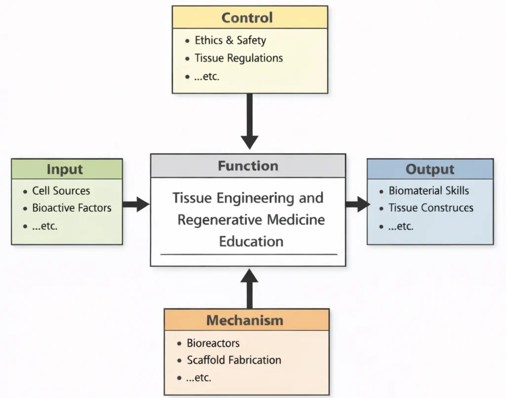
IDEF0 architectural model of tissue engineering education: biological inputs and signaling cues governed by regulatory controls, operationalized via automated bioreactor perfusion and precision scaffold additive manufacturing to yield clinical-grade functional constructs.
Tissue Engineering and Regenerative Medicine stands at the forefront of medical innovation, seeking to restore or replace biological tissues through the integration of biology, engineering, and material science. Closely associated with the broader field of Biomedical Engineering, it brings together tools from Bioinformatics to analyze cellular behaviors and optimize scaffold design. The development of biocompatible and biodegradable structures relies heavily on insights from Biomaterials, ensuring safety and functional integration within the human body.
Mechanical stimuli and load-bearing functions in engineered tissues are deeply informed by Biomechanics, while signal pathways that guide cell growth are deciphered using Biomedical Signal Processing. Applications within Cardiovascular Engineering include bioengineered blood vessels and heart valves, opening avenues to address congenital or degenerative cardiac conditions.
Clinical adoption of regenerative therapies depends on support from Clinical Engineering, which ensures compatibility with hospital systems and patient safety protocols. Meanwhile, diagnostic techniques developed in Medical Imaging are essential to visualize implanted tissues and monitor their integration over time. Research into neural tissue scaffolds also aligns with breakthroughs in Neural Engineering, where complex nerve regeneration is being made feasible.
Drug-eluting scaffolds and targeted tissue repair require close collaboration with experts in Pharmaceutical Engineering. For rehabilitation and prosthetics, engineered tissues are often integrated with adaptive devices designed in Rehabilitation Engineering. Multiscale tissue architecture must also be informed by foundational principles from Chemical Engineering and its numerous subdisciplines.
Production of cell culture media and bioreactors is optimized by methodologies from Biochemical Engineering. Reaction dynamics, especially in stem-cell signaling and scaffold-cell interactions, benefit from models derived from Chemical Catalysis and Reaction Engineering. For portable therapeutic devices, powering cell stimulation or on-site monitoring may require energy solutions developed in Chemical Energy Systems Engineering.
Advanced bioresorbable materials for tissue repair are engineered through Chemical Materials Engineering, while scaled-up production of tissue constructs involves principles from Chemical Process Engineering. Sophisticated simulations of tissue growth patterns and stress distributions are made possible by Computational Chemical Engineering. Certain applications, such as regenerating oral or gastrointestinal tissues, may intersect with challenges addressed in Food and Beverage Engineering.
Nano-engineered surfaces to guide cell differentiation or prevent infection are developed using tools from Nanotechnology in Chemical Engineering. The strength, flexibility, and degradability of scaffolds are customized through materials knowledge rooted in Polymer and Plastics Engineering. Supporting infrastructure for regenerative therapy centers is planned by professionals in Civil Engineering and constructed under guidance from Construction Management.
Finally, to maintain continuity of care during natural disasters or health emergencies, adaptive deployment strategies draw on Earthquake and Disaster Engineering. Altogether, Tissue Engineering and Regenerative Medicine embodies a confluence of life sciences, physical technologies, and innovation—offering hope for healing and human restoration in profoundly transformative ways.
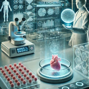
Convergence of hydrogel chemistry, stem cell differentiation pathways, and digital organ-scale bio-printing platforms within an advanced regenerative engineering facility.
This image portrays tissue engineering as a bridge between biology and engineering inside a high-tech laboratory. In the foreground, neatly arranged culture wells suggest cell growth and controlled experiments, while a microscope represents close observation of cells and tissue structure. At the center, a heart-like tissue construct sits on an illuminated platform, symbolizing lab-grown tissues and organ models used for repair, testing, or transplantation research. To the side, a transparent scaffold-like block hints at biomaterials and porous frameworks that support cell attachment and tissue formation. The researcher holding a glowing sphere evokes advanced design—such as engineered hydrogels, nanoparticles, or "smart" biomaterials—guided by digital measurements and biological data shown on surrounding screens. Overall, the illustration communicates the core idea of tissue engineering: designing living replacements by shaping cells and materials into functional structures that can restore damaged organs and improve human health.
Rebuilding Living Systems Through Engineering
Centres on reconstructing functional biological structures by guiding cells, materials, and biochemical cues into organised systems within the broader biomedical engineering roadmap.
Reveals genetic instructions and molecular pathways that determine how new tissues form, differentiate, and respond to environmental stimuli.
Delivers targeted signalling molecules, growth factor release systems, and therapeutics that guide cell behavior during active regeneration.
Provides tailor-made three-dimensional frameworks and synthetic matrix polymers that support cell attachment and guide structural organisation.
Shapes how physical stress, fluid shear, and mechanical forces influence tissue development, cellular alignment, and long-term functionality.
Tracks non-invasive structural formation, vascular integration, and host adaptation of engineered tissues inside living organisms over time.
Interprets bioelectric and functional signals to verify whether regenerated heart, muscle, or neural tissues are performing as intended.
Addresses restoration of neural communication pathways where regenerated tissues must successfully reconnect with central host networks.
Applies regenerative strategies to fabricate flow-capable biological constructs like vascular grafts, heart valves, and myocardial patches.
Integrates regenerated musculoskeletal tissue constructs into functional real-world recovery protocols and assistive biomechanical devices.
Oversees the safe translation, regulatory verification, and hospital deployment of advanced bioengineered tissue products.
Connects specific tissue engineering methodologies to the overarching discipline uniting engineering principles with life sciences.
Core Concepts in Tissue Engineering and Regenerative Medicine
Scaffold Design and Fabrication
Scaffolds serve as essential frameworks to guide the growth and regeneration of new tissues. Their three-dimensional (3D) architecture replicates the extracellular matrix (ECM), facilitating cell adhesion, proliferation, differentiation, and eventual integration into host tissues. Effective scaffold design involves a careful balance between porosity for nutrient transport and sufficient mechanical strength to sustain physiological stresses.
Governing dynamic relation: \(M_{\text{tot}}(t) = 100 e^{-k_{\text{deg}} t} + \frac{100 \varepsilon}{1 + e^{-r_{\text{ecm}}(t - 4)}}\). Online tool URL: https://prep4uni.online/stem/physical-technologies/biomedical-engineering/tissue-engineering-and-regenerative-medicine/
Materials in Scaffold Design
- Natural Polymers: Materials like collagen, chitosan, gelatin, and alginate closely mimic native ECM, presenting natural cell-adhesion ligands (e.g., Arg-Gly-Asp [RGD] sequences) while minimizing initial host inflammatory responses.
- Synthetic Polymers: Poly(\(\text{L}\)-lactic acid) (PLLA), polyglycolic acid (PGA), polycaprolactone (PCL), and polyethylene glycol (PEG) hydrogels provide controlled hydrolytic degradation rates, reproducible mechanical properties, and predictable thermoplastic processing.
Critical Properties of Scaffolds
- Biocompatibility & Immuno-Inertia: Must support cellular adhesion and remodeling without evoking severe foreign body reactions, fibrous encapsulation, or cytotoxic by-product release.
- Interconnected Porosity & Permeability: High open porosity (\(\varepsilon \ge 70\text{--}90\%\)) with pore diameters exceeding \(100\text{--}300\,\mu\text{m}\) is necessary for vascular ingrowth, mass convective transport of nutrients, and metabolic waste clearance.
- Mechanical Integrity & Viscoelastic Match: Scaffolds must withstand in vivo mechanical loading (e.g., \(1\text{--}20\,\text{GPa}\) for cortical/trabecular bone, \(0.5\text{--}5\,\text{MPa}\) for articular cartilage) without catastrophic collapse before nascent ECM can carry physiological shear and compressive loads.
Advanced Scaffold Fabrication Techniques
- 3D Bioprinting & Micro-Extrusion: Layer-by-layer spatial deposition of multi-cellular bio-inks and hydrogels, allowing micro-capillary channel patterning and spatially zoned mechanical stiffness.
- Electrospinning: Utilizing high-voltage electrostatic fields to draw polymer melts or solutions into sub-micron fibers (\(50\text{--}800\,\text{nm}\)), replicating the architectural topography and high surface area of native collagen fibrils.
Cell Sources and Their Applications
The selection of appropriate cell types is critical for successful tissue regeneration, influencing compatibility, integration, and long-term function.
Autologous Cells
- Derived directly from the patient, eliminating donor-to-host major histocompatibility complex (MHC) mismatch and graft rejection.
- Commonly used in bone marrow aspirates, autologous chondrocyte implantation (ACI), and split-thickness skin grafts, though restricted by donor site morbidity and patient age-dependent senescence.
Allogeneic Cells
- Sourced from human donors of the same species, facilitating cryopreserved, standardized, off-the-shelf therapeutic manufacturing.
- Employed in allogeneic bone grafts and mesenchymal stromal cell lines, requiring immunomodulatory co-treatments or human leukocyte antigen (HLA) screening.
Stem Cell Technologies
- Embryonic Stem Cells (ESCs): Pluripotent derivatives of the blastocyst inner cell mass with unlimited proliferative potential, subject to stringent ethical boundaries and teratoma formation risks.
- Adult Mesenchymal Stem/Stromal Cells (MSCs): Multipotent progenitors harvested from bone marrow, adipose tissue, or umbilical cord blood capable of osteogenic, chondrogenic, and adipogenic lineage differentiation.
- Induced Pluripotent Stem Cells (iPSCs): Somatic cells reprogrammed to pluripotency through the exogenous expression of Yamanaka factors (Oct4, Sox2, Klf4, c-Myc), providing patient-specific regenerative tissues and scalable pharmacological disease models.
Growth Factors and Biochemical Cues
Growth factors and biochemical signals orchestrate the cellular behaviors essential for successful tissue regeneration, directing cell migration, proliferation, and differentiation.
Role of Key Growth Factors
- Vascular Endothelial Growth Factor (VEGF): Potent angiogenic cytokine that stimulates endothelial cell proliferation, migration, and lumen formation to prevent ischemic necrosis in large constructs.
- Bone Morphogenetic Proteins (BMP-2, BMP-7): Transforming growth factor-beta (\(\text{TGF-}\beta\)) superfamily members that trigger runx2 gene transcription, driving osteogenic differentiation and mineralization.
- Fibroblast Growth Factors (FGF) & TGF-\(\beta\): Modulate fibroblast activation, collagen type I/III synthesis, and articular chondrocyte maintenance.
Delivery Strategies
- Controlled Release Systems: Nanosphere encapsulation, heparin-functionalized polymer matrices, and affinity hydrogels that sustain localized, zero-order release profiles, preventing the systemic toxicity of burst releases.
- Direct Local Injection: Administering concentrated growth factor solutions directly into localized lesions, providing transient stimulus but requiring high doses due to rapid in vivo protease degradation.
Bioreactors for Tissue Development
Bioreactors simulate physiological conditions, facilitating the controlled growth and maturation of engineered tissues in vitro.
Functions of Bioreactors
- Maintain homeostatic physiological environments through tight regulation of dissolved \(\text{O}_2\), \(\text{CO}_2\), pH (\(7.4 \pm 0.05\)), temperature (\(37^\circ\text{C}\)), and nutrient/metabolite mass transfer.
- Impart physiological mechanical conditioning: pulsatile luminal shear stress for vascular grafts, cyclic axial compression for articular cartilage constructs, and electrical pacing for engineered myocardium.
Types of Bioreactors
- Static Bioreactors: Culture vessels where mass transfer is driven solely by passive molecular diffusion; restricted to ultra-thin constructs (\(\lt 200\,\mu\text{m}\)).
- Perfusion Bioreactors: Force culture medium directly through the porous interconnects of a scaffold, eliminating boundary layer diffusion resistance and applying interstitial fluid shear stress.
- Rotating Wall Vessel & Spinner Flask Bioreactors: Provide microgravity-like low-shear environments or turbulent fluid mixing that supports cell seeding and spherical aggregate cultivation.
Immunological Considerations
Successful integration of engineered tissues requires careful management of immune responses to minimize rejection.
- Immune Modulation Techniques: Functionalization of biomaterial interfaces with anti-inflammatory molecules (e.g., IL-4, CD47 "don't-eat-me" signaling peptides) or hydrogel micro-encapsulation to isolate allogeneic cells from host cytotoxic T-lymphocytes.
- Personalized Approaches: Combining autologous cell sources or gene-edited hypoimmunogenic cell lines (e.g., CRISPR knockout of \(\text{B2M}\) to disrupt MHC Class I presentation) to minimize graft destruction.
Clinical Translation and Regulatory Pathways
Transitioning from laboratory research to clinical application involves rigorous testing and regulatory approval.
- Preclinical Testing: In vitro cytocompatibility assays (ISO 10993) followed by progressive animal studies (small rodent ectopic models up to large animal orthotopic implants) establishing safety, mechanical resilience, and integration.
- Regulatory Frameworks: Compliance with FDA Advanced Therapy Medicinal Product (ATMP) and Center for Biologics Evaluation and Research (CBER) rules (e.g., 21 CFR Part 1271 for HCT/Ps), or European Medicines Agency (EMA) Regulation (EC) No 1394/2007.
Contemporary 2026 AI-Era Computational Paradigms in Regenerative Medicine
Modern regenerative engineering integrates physical mechanics with deep learning frameworks to predict and optimize multi-scale tissue morphogenesis:
- Physics-Informed Neural Networks (PINNs) for Nutrient Transport: PINNs solve coupled Brinkman-Darcy momentum equations and Fickian convection-diffusion-reaction equations across complex 3D-bioprinted porous geometries without mesh generation:
$$\mathcal{L}_{\text{PINN}} = \left\| \nabla \cdot \mathbf{u} \right\|^2 + \left\| \frac{\mu}{\varepsilon} \nabla^2 \mathbf{u} - \nabla p - \frac{\mu}{K}\mathbf{u} \right\|^2 + \lambda \left\| \mathbf{u}\cdot\nabla C - D_{\text{eff}}\nabla^2 C + \frac{R_{\max} C}{K_m + C} \right\|^2$$
This allows rapid evaluation of hypoxic core formation in large tissue constructs under dynamic perfusion.Numerical Evaluation Example: Evaluating PINN Residuals Across a Perfused Scaffold Channel
To illustrate how a PINN evaluates physical residuals during backpropagation, consider an interstitial interrogation point inside a 3D-bioprinted porous osteochondral scaffold undergoing dynamic perfusion along the \(x\)-axis. The local physical and biological parameters are:
- Dynamic fluid viscosity: \(\mu = 1.0 \times 10^{-3}\,\text{Pa}\cdot\text{s}\)
- Scaffold permeability: \(K = 2.5 \times 10^{-11}\,\text{m}^2\)
- Scaffold porosity: \(\varepsilon = 0.70\)
- Effective oxygen diffusion coefficient: \(D_{\text{eff}} = 1.8 \times 10^{-9}\,\text{m}^2/\text{s}\)
- Maximum cellular metabolic oxygen consumption: \(R_{\max} = 1.2 \times 10^{-4}\,\text{mol/(m}^3\cdot\text{s)}\)
- Michaelis-Menten constant: \(K_m = 0.010\,\text{mol/m}^3\)
At collocation coordinate \((x^*, y^*, z^*)\), the automatic differentiation (AD) engine evaluates the network output variables and their spatial gradients:
- Local fluid velocity: \(u_x = 1.5 \times 10^{-4}\,\text{m/s}\) (with \(\nabla \cdot \mathbf{u} \approx 0\))
- Local pressure gradient: \(\frac{\partial p}{\partial x} = -6.05 \times 10^3\,\text{Pa/m}\)
- Laplacian of velocity: \(\nabla^2 u_x = -1.2 \times 10^4\,\text{m}^{-1}\cdot\text{s}^{-1}\)
- Local oxygen concentration: \(C = 0.040\,\text{mol/m}^3\)
- Concentration axial gradient: \(\frac{\partial C}{\partial x} = -18.5\,\text{mol/m}^4\)
- Concentration Laplacian: \(\nabla^2 C = 3.6 \times 10^4\,\text{mol/m}^5\)
Step 1: Evaluate the Brinkman-Darcy Momentum Residual (\(r_{\text{mom}}\))
$$r_{\text{mom}} = \frac{\mu}{\varepsilon} \nabla^2 u_x - \frac{\partial p}{\partial x} - \frac{\mu}{K} u_x$$ $$\begin{aligned} \frac{\mu}{\varepsilon} \nabla^2 u_x &= \frac{1.0 \times 10^{-3}}{0.70} \times (-1.2 \times 10^4) = -17.14\,\text{Pa/m} \\ -\frac{\partial p}{\partial x} &= -(-6.05 \times 10^3) = +6050.0\,\text{Pa/m} \\ -\frac{\mu}{K} u_x &= -\frac{1.0 \times 10^{-3}}{2.5 \times 10^{-11}} \times (1.5 \times 10^{-4}) = -(4.0 \times 10^7) \times (1.5 \times 10^{-4}) = -6000.0\,\text{Pa/m} \end{aligned}$$ $$r_{\text{mom}} = -17.14 + 6050.0 - 6000.0 = \mathbf{+32.86\,\text{Pa/m}}$$
Step 2: Evaluate the Fickian Convective-Diffusive-Reactive Nutrient Residual (\(r_{\text{nutr}}\))
$$r_{\text{nutr}} = u_x \frac{\partial C}{\partial x} - D_{\text{eff}} \nabla^2 C + \frac{R_{\max} C}{K_m + C}$$ $$\begin{aligned} \text{Convective term: } u_x \frac{\partial C}{\partial x} &= (1.5 \times 10^{-4}\,\text{m/s}) \times (-18.5\,\text{mol/m}^4) = -2.775 \times 10^{-3}\,\text{mol/(m}^3\cdot\text{s)} \\ \text{Diffusive term: } -D_{\text{eff}} \nabla^2 C &= -(1.8 \times 10^{-9}\,\text{m}^2/\text{s}) \times (3.6 \times 10^4\,\text{mol/m}^5) = -6.48 \times 10^{-5}\,\text{mol/(m}^3\cdot\text{s)} \\ \text{Metabolic uptake: } \frac{R_{\max} C}{K_m + C} &= \frac{(1.2 \times 10^{-4}) \times (0.040)}{0.010 + 0.040} = \frac{4.8 \times 10^{-6}}{0.050} = +9.60 \times 10^{-5}\,\text{mol/(m}^3\cdot\text{s)} \end{aligned}$$ $$r_{\text{nutr}} = -2.775 \times 10^{-3} - 6.48 \times 10^{-5} + 9.60 \times 10^{-5} = \mathbf{-2.744 \times 10^{-3}\,\text{mol/(m}^3\cdot\text{s)}}$$
Physical & Computational Interpretation: The non-zero residuals (\(r_{\text{mom}} = 32.86\,\text{Pa/m}\) and \(r_{\text{nutr}} = -2.744 \times 10^{-3}\,\text{mol/(m}^3\cdot\text{s)}\)) represent the exact local physics violations passed to the optimizer via automatic differentiation. By squaring and minimizing these residuals across thousands of unmeshed spatial coordinates simultaneously, the PINN converges toward exact mass, momentum, and biological conservation, predicting whether the downstream core falls below the critical hypoxia boundary (\(C \lt 0.010\,\text{mol/m}^3\)) without needing a boundary-fitted computational grid.
- DeepONets & Neural Operators for Scaffold Remodeling: Deep Operator Networks learn the non-linear mappings between patient-specific musculoskeletal loading cycles and spatial bio-degradation dynamics, enabling real-time optimization of 3D print architectures.
- Generative Foundation Models for Bio-Ink Rheology: Generative models trained on polymer cross-linking kinetics predict the printability window (viscosity, yield stress, loss tangent \(\tan\delta\)) of multi-material hydrogel suspensions, minimizing pre-extrusion cell mechanical lysis.
Future Directions in Tissue Engineering
- 4D Bioprinting: Producing constructs using stimuli-responsive smart materials (e.g., shape-memory hydrogels) that dynamically alter their geometric configurations in response to temperature, hydration, or cellular traction forces.
- Advanced Computational Modeling & In Silico Trials: Digital twins of bioreactors simulating cell proliferation kinetics, oxygen gradients, and flow shear stress to optimize manufacturing parameters before physical cultivation.
Ethical and Societal Implications
- Ethical Frameworks: Formulating international standards concerning human embryonic stem cells, chimera research, and the potential editing of germline-adjacent regenerative systems.
- Public Engagement and Policy: Establishing socio-economic mechanisms to ensure that personalized regenerative therapies and lab-grown organs are equitably accessible rather than restricted to high-income settings.
Major Techniques in Tissue Engineering and Regenerative Medicine
3D Bioprinting
- Definition: Additive biofabrication depositing spatial arrays of living cells, biochemical factors, and biocompatible hydrogels in a computer-directed layer-by-layer sequence.
- Applications:
- Fabricating full-thickness cellular skin grafts, osteochondral plugs, and branched vascular trees.
- Constructing microfluidic organ-on-a-chip disease models for pharmaceutical screening.
- Advantages:
- High geometric precision matching patient-specific DICOM computed tomography datasets.
- Direct co-deposition of multiple distinct cell lineages and specialized extracellular matrices.
Decellularization and Recellularization
- Definition: Chemical, physical, or enzymatic stripping of all antigenic cellular components from a donor organ, leaving an intact extracellular matrix architecture that is subsequently re-seeded with recipient-matched cells.
- Applications:
- Bioengineered cardiac valves, tracheobronchial scaffolds, and whole porcine renal scaffolds.
- Perfusion recellularization of hepatic and pulmonary organ frameworks.
- Advantages:
- Preserves native microvascular topology, basement membrane proteins, and anatomical architecture.
- Significantly reduces hyperacute host immunogenicity compared to untreated xenografts.
Stem Cell Therapy
- Definition: Direct transplantation of multipotent or pluripotent stem cells to accelerate endogenous tissue repair through differentiation or paracrine trophic signaling.
- Applications:
- Mesenchymal stromal cell (MSC) injection for osteoarthritis and critical-sized bone defects.
- Neural stem cell grafting for spinal cord transsection lesions.
- Advantages:
- High proliferative capacity and broad multipotent differentiation potential.
- Robust secretion of regenerative exosomes, immunomodulatory cytokines, and angiogenic factors.
Gene Editing and Gene Therapy
- Definition: Precision modification of chromosomal sequences within reparative cells to suppress disease alleles, enhance survival, or overexpress pro-regenerative cytokines.
- Technologies:
- CRISPR-Cas9, base editing, and prime editing systems delivering targeted genomic modifications.
- Recombinant adeno-associated viruses (rAAV) and synthetic lipid nanoparticles for non-viral transduction.
- Applications:
- Knocking out \(\text{HLA}\) surface loci to generate universal allogeneic stem cell lines.
- Engineering cartilage grafts resistant to systemic inflammatory cytokines (\(\text{IL-1}\beta\), \(\text{TNF-}\alpha\)).
Applications of Tissue Engineering and Regenerative Medicine
Organ Regeneration and Transplantation
- Bioengineered Organs: Laboratory development of functional renal filtration units, vascularized cardiac patches, and hepatic lobule constructs using perfusion bioreactors.
- Impact: Alleviates the global shortage of donor organs, reducing mortality on clinical waiting lists.
Skin Tissue Engineering
- Applications: Living dermo-epidermal bilayer replacements for extensive third-degree burn wounds, chronic diabetic foot ulcers, and reconstructive plastic surgery.
- Materials Used: Cross-linked type I collagen sponges, hyaluronic acid hydrogels, and bioprinted autologous keratinocyte/fibroblast constructs.
Cartilage and Bone Regeneration
- Cartilage Repair: Avascular hydrogel frameworks (alginate, agarose, chondroitin sulfate) seeded with chondrocytes to repair articular cartilage defects without fibrocartilage scarring.
- Bone Regeneration: Highly porous bioactive glass (\(45\text{S}5\)), hydroxyapatite, and beta-tricalcium phosphate (\(\beta\text{-TCP}\)) ceramic matrices for orthopedic critical defect repair.
Cardiovascular Tissue Engineering
- Applications: Tissue-engineered vascular grafts (TEVGs) exhibiting physiological burst pressures, compliant bi-leaflet heart valves, and contractility-synchronized cardiomyocyte patches.
- Materials Used: Decellularized arterial conduits and biodegradable synthetic polymers (e.g., polyglycolic acid woven meshes).
Nerve and Spinal Cord Regeneration
- Applications: Engineered neural guidance conduits (NGCs) bridging peripheral nerve gaps (\(\gt 30\,\text{mm}\)) and conductive hydrogel matrices for spinal cord repair.
- Materials: Aligned electrospun PCL nanofibers, conductive polymers (polypyrrole, PEDOT:PSS), and laminin functional coatings.
Drug Testing and Disease Modeling
- Organ-on-a-Chip: Microfluidic devices integrating cellular co-cultures under continuous micro-perfusion, replicating mechanical and biochemical organ interfaces.
- 3D Tissue Models: High-throughput human disease screens that reduce dependence on animal testing while enhancing predictive pharmacological efficacy and toxicology screening.
Challenges in Tissue Engineering and Regenerative Medicine
Vascularization
Creating functional, hierarchically branched vascular networks remains a primary constraint for constructs thicker than the diffusion limit of oxygen (\(\sim 150\text{--}200\,\mu\text{m}\)). Cells residing within the core of larger unvascularized constructs experience hypoxia, localized acidosis, and necrosis. While sacrificial carbohydrate glass printing and 3D bioprinting of endothelialized microfluidic channels have improved outcomes, achieving seamless microvascular anastomosis with the host circulation upon implantation remains an active area of investigation.
Immune Rejection
Engineered tissues and implanted biomaterials frequently elicit immune responses that hinder therapeutic outcomes. Foreign cells or synthetic scaffolds can activate inflammation, fibrosis, or immune rejection, limiting long-term viability. Strategies like immunosuppressive therapies and genetic modification of cells to evade immune detection have shown potential but carry significant risks and complexity. Allogeneic (donor-derived) tissue engineering products face higher risks of rejection compared to autologous (patient-derived) materials, complicating their clinical adoption. Development of immune-compatible or immune-evasive biomaterials and scaffolds is essential for reducing rejection. Advanced stem cell techniques, including induced pluripotent stem cells (iPSCs), offer promise in personalized medicine, potentially mitigating rejection issues. Understanding immune mechanisms involved in rejection at the cellular and molecular levels remains a crucial research area to enhance clinical outcomes.
Mechanical Strength
Replicating the mechanical properties of native tissues—such as strength, elasticity, and toughness—is a persistent challenge, particularly for load-bearing tissues like bone, cartilage, and tendons. Most engineered tissues currently lack the biomechanical resilience and durability of their natural counterparts, limiting clinical applications. Innovations in composite biomaterials, incorporating natural proteins (collagen, elastin) and synthetic polymers, are being developed to enhance mechanical integrity. Biomechanical testing methods and computational simulations play critical roles in predicting and validating mechanical performance. Long-term durability and fatigue resistance are essential yet difficult to achieve consistently in tissue-engineered constructs. Emerging techniques such as hybrid scaffolds with reinforcing fibers, nanoparticle reinforcement, and bioactive glass materials are being explored for improved strength.
Integration with Host Tissues
Ensuring seamless integration between engineered tissues and host tissues is crucial for long-term success and functionality. Poor integration can result in implant failure, impaired tissue regeneration, or fibrosis around the implant site. Developing scaffold materials that closely mimic the native extracellular matrix (ECM) supports improved integration and tissue remodeling. Advanced surface modifications, bioactive coatings, and growth factor delivery systems aim to facilitate cellular infiltration and integration. Controlled biodegradation rates of scaffolds are critical to match the natural tissue regeneration timeline, supporting gradual integration and remodeling. Preclinical animal models are widely used to assess integration, but these results often differ from clinical realities.
Regulatory and Ethical Challenges
Tissue-engineered products involve complex combinations of cells, biomaterials, and biological factors, posing substantial regulatory challenges. Stringent clinical testing, validation, and safety assessments required by agencies like the U.S. Food and Drug Administration (FDA) and the European Medicines Agency (EMA) significantly extend development timelines and costs. Standardizing processes and quality control methods for tissue-engineered products remains difficult due to biological variability and manufacturing complexity. Ethical considerations around the sourcing and use of human stem cells, particularly embryonic stem cells, continue to prompt debate and scrutiny. Informed consent, patient privacy, and transparency in clinical trials are essential ethical concerns that must be managed effectively. Robust regulatory frameworks and ethical guidelines are crucial for public trust and widespread clinical adoption of regenerative therapies.
Scalability and Manufacturing
Transitioning from laboratory-scale research to large-scale commercial production of engineered tissues is a major challenge. Manufacturing processes must be standardized, reproducible, cost-effective, and capable of mass production while maintaining biological efficacy. Automation, bioreactors, and advanced manufacturing technologies, including 3D bioprinting, are under investigation to improve scalability. Quality assurance and process validation at large scale requires innovative methods to ensure consistent cell viability, functionality, and scaffold properties. Supply chain management, particularly the consistent sourcing of raw materials and biological components, poses additional scalability challenges.
Cell Source and Quality Control
The choice and quality of cellular sources significantly affect the success of engineered tissues. Challenges include variability in stem cell differentiation, maintaining cellular phenotype, and controlling genetic stability during expansion. Reliable quality control assays and standardization of cell culture practices are vital for ensuring consistent cellular behavior. Emerging developments in cell reprogramming and genetic engineering technologies offer potential solutions but introduce new complexities and regulatory considerations. Advanced cell banking strategies and comprehensive characterization protocols are essential to improve reliability and reproducibility.
Further Reading and External Resources:
- National Institute of Biomedical Imaging and Bioengineering (NIBIB) – Comprehensive resource on tissue engineering and regenerative medicine research and applications.
- International Society for Stem Cell Research (ISSCR) – Global standards, ethical guidelines, and research updates on stem cells and regenerative medicine.
Emerging Trends and Future Directions in Tissue Engineering and Regenerative Medicine
4D Bioprinting: Dynamic and Responsive Tissue Constructs
- Definition and Principle: 4D bioprinting refers to the creation of tissue constructs capable of changing shape, structure, or function over time in response to environmental stimuli such as temperature, moisture, pH, or mechanical forces.
- Materials and Mechanisms: Smart biomaterials including shape-memory polymers, hydrogels, and responsive nanocomposites are engineered to exhibit controlled responses.
- Potential Applications: Vascular grafts that expand post-implantation, responsive drug delivery systems, and dynamic implants for repairing organs like the heart and lungs.
- Research Advances: Recent studies have demonstrated the feasibility of printed scaffolds that self-fold or expand upon exposure to bodily fluids, significantly enhancing tissue regeneration capabilities.
- Challenges and Future Prospects: Further developments are necessary to improve reliability, predictability, and scalability of these responsive systems for clinical translation.
Organoids and Personalized Medicine
- Definition and Importance: Organoids are miniature, three-dimensional, lab-grown tissues that replicate organ-specific structures and functions, derived from stem cells or patient-specific cells.
- Applications in Disease Modeling: Used to mimic human diseases at a microscopic scale, enabling precise studies of pathology, genetic mutations, and therapeutic responses.
- Drug Testing and Development: Provide powerful models for high-throughput drug screening, personalized therapy assessments, and reducing reliance on animal testing.
- Clinical Relevance: Patient-derived organoids help tailor treatments for diseases like cancer, cystic fibrosis, and inflammatory bowel disease, significantly advancing personalized medicine.
- Challenges in Standardization: Variability in organoid growth and maturation necessitates development of standardized protocols and validation methods for consistent outcomes.
- Future Directions: Integration of organoid technology with advanced imaging, microfluidics, and artificial intelligence (AI) to further enhance accuracy and predictive capabilities.
Synthetic Biology in Tissue Regeneration
- Concept and Goals: Synthetic biology involves the engineering of novel biological systems to enhance or replicate tissue regeneration processes.
- Genetic Engineering: Modifying genetic pathways in cells to promote rapid healing, controlled growth, or enhanced functional integration of tissues.
- Programmable Cells: Developing genetically engineered stem cells capable of sensing environmental cues and responding accordingly to enhance regeneration and integration.
- Biofabrication Techniques: Synthetic biology-driven approaches to create scaffolds with enhanced biological functionality, including embedded growth factors and bioactive signals.
- Ethical and Regulatory Considerations: Requires careful evaluation of biosafety, ethical implications, and long-term impacts before widespread clinical application.
- Future Outlook: Promising research is underway to harness synthetic biology for advanced regenerative therapies, potentially revolutionizing medicine in the next decade.
Integration with Wearable Devices and Biosensors
- Combined Technologies: Emerging trend in integrating tissue-engineered constructs with wearable or implantable biosensors for continuous monitoring of tissue health and physiological functions.
- Real-Time Monitoring: Devices capable of detecting biomarkers, tissue integrity, inflammation, or infection early, facilitating proactive clinical interventions.
- Examples and Research: Development of smart dressings that monitor wound healing or implantable scaffolds equipped with sensors for real-time feedback on regeneration progress.
- Data Integration and Analysis: Advanced algorithms, machine learning, and AI platforms analyze sensor data, enabling personalized and timely clinical decisions.
- Technical Challenges: Ensuring sensor biocompatibility, miniaturization, wireless data transmission, and long-term reliability in physiological environments.
- Future Potential: Pioneering efforts are expected to yield next-generation regenerative implants with sophisticated, integrated diagnostic capabilities.
Large-Scale Organ Manufacturing
- Concept and Need: Large-scale, clinical-grade manufacturing of functional, transplantable organs addresses critical organ shortages for transplants.
- Technologies Involved: Advanced 3D bioprinting, automated cell culturing systems, and tissue maturation bioreactors are central to this effort.
- Biofabrication Progress: Current developments demonstrate feasibility in producing functional tissues like skin, cartilage, and vascular grafts, with ongoing research aimed at more complex organs such as kidneys and hearts.
- Scalability Challenges: Significant hurdles include reliable vascularization, maintaining organ viability and function post-fabrication, and meeting regulatory standards.
- Ethical and Social Considerations: Accessibility, cost-effectiveness, and ethical questions surrounding large-scale human organ production must be addressed for societal acceptance.
- Potential Impacts: Successful clinical translation would significantly reduce organ donation waiting lists, dramatically changing healthcare landscapes.
Bioinformatics and Computational Modeling
- Integrating bioinformatics tools and computational models to optimize scaffold design, predict cellular behavior, and improve experimental outcomes in tissue engineering.
- Machine learning algorithms and big data analytics increasingly guide experimental strategies and scaffold customization.
Stem Cell Engineering Innovations
- Advances in induced pluripotent stem cells (iPSCs) and direct lineage reprogramming enable highly personalized regenerative treatments.
- Efficient differentiation protocols to generate specific tissue types continue to improve, enhancing clinical feasibility and precision.
Extracellular Matrix (ECM) Mimicry
- Innovative biomaterials closely mimicking native ECM properties enhance cell growth, differentiation, and tissue regeneration outcomes.
- Biochemical and mechanical cues engineered into scaffolds better replicate native tissue environments, improving integration and functionality.
Further Reading and Resources:
- Wake Forest Institute for Regenerative Medicine – Leading research institute pioneering clinical translation of regenerative therapies.
- Nature - Tissue Engineering Research – Comprehensive collection of latest research, reviews, and advancements in tissue engineering and regenerative medicine.
Systems Engineering Trade-Off Analysis in Regenerative Scaffold Design
Designing regenerative scaffolds involves balancing conflicting biological, mechanical, and transport requirements. The following matrix illustrates key engineering compromises:
| Design Domain | Parameter Dimension A | Parameter Dimension B | Critical Multi-Variable Compromise |
|---|---|---|---|
| Scaffold Architecture | High Porosity & Large Pore Diameter (\(\varepsilon \gt 85\%\), \(d_{\text{pore}} \gt 300\,\mu\text{m}\)) | Compressive Modulus & Yield Strength (\(E \gt 15\,\text{GPa}\) for bone) | Maximizing open volume for cell infiltration and vascularization versus maintaining structural integrity under physiological skeletal loading. |
| Degradation Kinetics | Rapid Hydrolytic Resorption (e.g., High PGA content: \(t_{1/2} \approx 2\text{--}4\,\text{weeks}\)) | Extended Biomechanical Support (e.g., PCL / PLLA: \(t_{1/2} \gt 1\text{--}2\,\text{years}\)) | Preventing long-term foreign body response versus premature load transfer causing graft collapse before endogenous ECM mineralizes. |
| Bio-Ink Rheology | High Viscosity & Crosslinking Density (High Shape Fidelity) | Low Extrusion Shear Stress (\(\tau \lt 5\,\text{kPa}\) for cell viability) | Ensuring 3D printed microstructures do not collapse post-deposition versus preventing shear-induced cell membrane lysis during needle passage. |
| Cellular Sourcing | Autologous Primary Isolates (Zero MHC rejection) | Allogeneic / iPSC Off-the-Shelf Banks (Immediate scalability) | Eliminating chronic immunosuppression versus manufacturing delays (weeks/months) and age-associated donor cell senescence. |
Worked Engineering Calculation: Scaffold Core Hypoxia & Critical Thickness Limit
Analytical Engineering Derivation: Critical Diffusion Dimension in Engineered Constructs
Problem Formulation: An unvascularized flat slab tissue-engineered construct of total thickness \(2L\) is seeded uniformly with cells at high density (\(\rho_{\text{cell}} = 2.5 \times 10^7\,\text{cells/cm}^3\)). Both top and bottom faces (\(x = \pm L\)) are exposed to well-oxygenated culture medium maintaining a surface oxygen concentration of \(C_0 = 0.20\,\text{mol/m}^3\). The cells consume oxygen following zero-order kinetics at an individual rate of \(q_{\text{cell}} = 1.0 \times 10^{-17}\,\text{mol/(cell}\cdot\text{s)}\), giving a volumetric oxygen consumption rate \(Q = \rho_{\text{cell}} \times q_{\text{cell}}\). The effective diffusion coefficient of oxygen through the cell-seeded hydrogel matrix is \(D_{\text{eff}} = 1.6 \times 10^{-9}\,\text{m}^2/\text{s}\).
- Calculate the volumetric consumption rate \(Q\) in \(\text{mol/(m}^3\cdot\text{s)}\).
- Derive the analytical expression for the steady-state oxygen concentration profile \(C(x)\) across the slab using Fick's second law with consumption.
- Determine the maximum allowable half-thickness \(L_{\text{crit}}\) (and total thickness \(2L_{\text{crit}}\)) of the construct before the core concentration drops below the anoxic necrosis threshold (\(C(0) = 0\,\text{mol/m}^3\)).
Step 1: Calculate volumetric consumption rate \(Q\)
$$\begin{aligned}
\rho_{\text{cell}} &= 2.5 \times 10^7\,\frac{\text{cells}}{\text{cm}^3} = 2.5 \times 10^{13}\,\frac{\text{cells}}{\text{m}^3} \\
q_{\text{cell}} &= 1.0 \times 10^{-17}\,\frac{\text{mol}}{\text{cell}\cdot\text{s}} \\
Q &= (2.5 \times 10^{13}\,\text{cells/m}^3) \times (1.0 \times 10^{-17}\,\text{mol/(cell}\cdot\text{s)}) = \mathbf{2.5 \times 10^{-4}\,\frac{\text{mol}}{\text{m}^3\cdot\text{s}}}
\end{aligned}$$
Step 2: Solve governing 1D steady-state diffusion-reaction equation
The 1D mass balance across the construct is governed by:
$$D_{\text{eff}} \frac{\mathrm{d}^2C}{\mathrm{d}x^2} - Q = 0 \quad \Rightarrow \quad \frac{\mathrm{d}^2C}{\mathrm{d}x^2} = \frac{Q}{D_{\text{eff}}}$$
Integrating twice:
$$\begin{aligned}
\frac{\mathrm{d}C}{\mathrm{d}x} &= \frac{Q}{D_{\text{eff}}} x + B_1 \\
C(x) &= \frac{Q}{2 D_{\text{eff}}} x^2 + B_1 x + B_2
\end{aligned}$$Applying symmetry boundary condition at the center (\(x = 0\)):
$$\left.\frac{\mathrm{d}C}{\mathrm{d}x}\right|_{x=0} = 0 \quad \Rightarrow \quad B_1 = 0$$Applying surface concentration boundary condition at \(x = L\):
$$C(L) = C_0 \quad \Rightarrow \quad C_0 = \frac{Q L^2}{2 D_{\text{eff}}} + B_2 \quad \Rightarrow \quad B_2 = C_0 - \frac{Q L^2}{2 D_{\text{eff}}}$$Thus, the spatial concentration profile is:
$$C(x) = C_0 - \frac{Q}{2 D_{\text{eff}}} \left( L^2 - x^2 \right)$$
At the construct mid-plane (\(x = 0\)):
$$C(0) = C_0 - \frac{Q L^2}{2 D_{\text{eff}}}$$
Step 3: Calculate critical half-thickness \(L_{\text{crit}}\)
Setting core concentration to zero (\(C(0) = 0\)):
$$0 = C_0 - \frac{Q L_{\text{crit}}^2}{2 D_{\text{eff}}} \quad \Rightarrow \quad L_{\text{crit}} = \sqrt{\frac{2 D_{\text{eff}} C_0}{Q}}$$Substitute empirical values:
$$\begin{aligned}
L_{\text{crit}} &= \sqrt{\frac{2 \times (1.6 \times 10^{-9}\,\text{m}^2/\text{s}) \times (0.20\,\text{mol/m}^3)}{2.5 \times 10^{-4}\,\text{mol/(m}^3\cdot\text{s)}}} \\
&= \sqrt{\frac{6.4 \times 10^{-10}}{2.5 \times 10^{-4}}} = \sqrt{2.56 \times 10^{-6}\,\text{m}^2} = \mathbf{1.60 \times 10^{-3}\,\text{m}} = \mathbf{1.60\,\text{mm}}
\end{aligned}$$
Total critical slab thickness:
$$2 L_{\text{crit}} = 2 \times 1.60\,\text{mm} = \mathbf{3.20\,\text{mm}}$$
Physical Interpretation & Design Implication:
If the construct half-thickness exceeds \(1.60\,\text{mm}\) (or \(\sim 160\text{--}200\,\mu\text{m}\) in spherical cell spheroids where diffusion drops along three axes), the core region will become completely anoxic, triggering cellular necrosis. This analytical boundary defines the maximum allowable thickness for passive diffusion culture, demonstrating why dynamic microvascular perfusion networks or bioprinted sacrificial channels are strictly required for clinical-scale human tissue regeneration.
Why Study Tissue Engineering and Regenerative Medicine
This field aims to create living tissues to replace damaged or diseased organs. Students learn to combine cells, scaffolds, and growth factors to support regeneration. These advances offer hope for treating previously incurable conditions.
Students study how to manipulate stem cells to form desired tissue types. This includes differentiation, signaling pathways, and genetic modification. These skills are essential for producing functional and targeted tissues.
The design of biocompatible scaffolds is crucial for supporting cell growth. Students learn how to fabricate materials that mimic natural tissue environments. This ensures proper structure, integration, and healing.
Tissue engineering has applications in wound healing, bone regeneration, and organ replacement. Students explore how lab-grown tissues can be implanted into patients. This bridges lab research with clinical practice.
Students are encouraged to innovate in bioprinting, immunomodulation, and large-scale production. The field faces challenges in vascularization and integration. Overcoming these barriers will redefine the future of medicine.
Tissue Engineering and Regenerative Medicine - Conclusion
A Transformative Approach to Healthcare
Tissue engineering and regenerative medicine stand at the forefront of healthcare innovation, fundamentally transforming approaches to treatment and recovery. By harnessing cutting-edge techniques to repair, regenerate, and replace damaged tissues, these disciplines promise not just symptomatic relief, but lasting solutions to previously untreatable conditions. The development of sophisticated biotechnologies such as biomaterial scaffolds, advanced stem cell applications, and innovative bioprinting methods underscores their role as a transformative force in modern medicine.
Advancements in 3D Bioprinting and Customization
The advent of 3D bioprinting represents one of the most groundbreaking advancements in regenerative medicine. This technology enables precise, patient-specific tissue constructs designed to match complex anatomical structures, significantly enhancing their integration and functionality post-implantation. Bioengineered tissues such as skin, cartilage, and blood vessels are already making significant clinical impacts, dramatically improving patient outcomes in burns, orthopedic injuries, and cardiovascular repair.
Stem Cells as Pioneers of Regeneration
Stem cell research remains central to the growth and potential of regenerative medicine. The unique ability of stem cells—particularly induced pluripotent stem cells (iPSCs)—to differentiate into various tissue types provides unprecedented opportunities to regenerate damaged tissues and restore organ functions. Clinical trials leveraging stem cells are showing promising results in treatments for conditions such as spinal cord injuries, heart disease, diabetes, and neurodegenerative disorders, highlighting their immense therapeutic potential.
Innovative Biomaterials and Scaffold Design
The development of innovative biomaterials plays a crucial role in tissue engineering, providing essential structural and biochemical support for cell growth and tissue regeneration. Advanced biomaterials mimic the extracellular matrix (ECM) environment, promoting cellular adhesion, proliferation, and differentiation. Scaffold technologies now incorporate bioactive molecules and growth factors, significantly enhancing tissue formation and integration, ultimately improving clinical outcomes for patients undergoing regenerative treatments.
Addressing Critical Healthcare Challenges
Tissue engineering and regenerative medicine directly address critical challenges such as organ shortages, chronic disease management, and complex injury recovery. Bioengineered organ substitutes and tissue grafts offer viable alternatives to traditional organ transplantation, reducing dependency on donor availability and minimizing immune rejection risks. These advancements promise to significantly alleviate global healthcare burdens, enhancing quality of life and extending lifespan for countless individuals.
Personalized and Precision Medicine
The intersection of regenerative medicine with precision medicine has fostered a highly personalized approach to treatment. Customized tissue-engineered products tailored to individual genetic profiles and anatomical requirements ensure more effective and safer therapeutic outcomes. As personalized medicine advances, the integration of genetic diagnostics, bioinformatics, and regenerative technologies will further optimize patient-specific treatments, transforming healthcare delivery into a highly individualized experience.
Multidisciplinary Collaboration for Accelerated Progress
Continued innovation in tissue engineering and regenerative medicine depends heavily on interdisciplinary collaboration. Biomedical engineers, material scientists, geneticists, clinicians, and bioinformatics experts are increasingly working together to accelerate research and translate laboratory breakthroughs into viable clinical therapies. These collaborative efforts facilitate rapid advancements, ensuring that innovative solutions are effectively developed, tested, and implemented into mainstream healthcare practices.
Overcoming Remaining Challenges
Despite significant progress, several challenges remain, including vascularization of larger engineered tissues, long-term immune compatibility, and regulatory hurdles for clinical translation. Addressing these issues requires ongoing research, development, and rigorous clinical trials. Continued investment in infrastructure, education, and regulatory frameworks is essential for overcoming these barriers and ensuring successful integration of regenerative technologies into widespread medical practice.
Ethical Considerations and Regulatory Frameworks
As tissue engineering evolves, ethical considerations surrounding stem cell use, genetic modifications, and large-scale tissue manufacturing remain critical areas of discussion. Developing comprehensive ethical guidelines and robust regulatory standards will be crucial to safeguarding patient safety and ensuring responsible innovation. Transparent dialogue among researchers, policymakers, healthcare providers, and the public will help address concerns and foster trust in regenerative therapies.
The Future Outlook
Looking forward, the future of tissue engineering and regenerative medicine is exceedingly promising. Advancements in artificial intelligence, machine learning, and computational modeling are poised to further revolutionize these fields by enhancing predictive capabilities, streamlining development processes, and accelerating clinical translation. Ultimately, continued innovation will make regenerative solutions increasingly accessible and affordable, transforming healthcare on a global scale and improving the lives of millions.
Tissue Engineering and Regenerative Medicine – Frequently Asked Questions (FAQ)
1. Is tissue engineering the same as "growing organs in the lab"?
Tissue engineering does include attempts to grow complex organs, but it also covers simpler constructs such as skin grafts, cartilage patches and bone scaffolds. Many clinically used products are currently small pieces of tissue rather than whole organs, yet they already play an important role in repair and reconstruction.
2. How is regenerative medicine broader than tissue engineering?
Regenerative medicine uses any strategy that helps the body repair itself, including stem cell injections, gene therapies and biologically active molecules that stimulate natural healing. Tissue engineering is one powerful tool inside this wider toolbox, focusing on building replacement tissues with cells and scaffolds.
3. Do all tissue-engineered constructs rely on stem cells?
Not always. Some constructs use differentiated cells taken from the patient, while others rely on stem cells or progenitor cells that can become many different tissue types. Stem cells are especially useful when large numbers of cells or specialised cell types are needed for regeneration.
4. Why are scaffolds necessary if cells can grow on their own?
In the body, cells rarely exist in isolation; they are supported by a complex extracellular matrix. Scaffolds mimic this environment, giving cells a place to attach and organise, and providing mechanical strength while new tissue forms. Without an appropriate scaffold, cells may not assemble into a stable, functional structure.
5. Can 3D bioprinting really produce usable human organs?
3D bioprinting is already used to create small tissue patches and detailed models of organs for research and drug testing. Printing full-sized, fully functional transplantable organs remains a long-term goal, mainly because of the difficulty of creating robust blood vessel networks and long-term function. However, progress is steady, and intermediate applications are already valuable.
6. What makes it so hard to move tissue-engineered products from lab to clinic?
Laboratory prototypes often work under carefully controlled conditions. Translating them to real patients requires proving safety, durability and reproducible manufacturing. Engineers must design processes that can be scaled up, meet stringent regulatory standards and still remain affordable for healthcare systems.
7. Will tissue engineering eliminate the need for organ donors?
In the near term, tissue engineering is more likely to complement organ donation rather than replace it. Engineered tissues can reduce the demand for certain transplants and improve outcomes, but creating complex, fully functional organs at scale is still an active area of research.
8. Are there ethical concerns with using stem cells in regenerative medicine?
Yes. Questions about how stem cells are obtained, how informed consent is handled and how experimental treatments are marketed all need careful attention. Many current approaches use adult or induced pluripotent stem cells, which avoid some of the ethical concerns associated with embryonic sources.
9. How does tissue engineering support personalised medicine?
Researchers can grow mini-tissues from a patient's own cells and test different drugs or treatment strategies on them before treating the patient. This helps predict which therapies are most likely to work and reduces the risk of serious side effects, making treatment more tailored and precise.
10. What background do students typically need to study tissue engineering?
Undergraduate preparation usually includes biology, chemistry, physics and mathematics, together with introductory courses in materials science and engineering. At university level, students may specialise through biomedical engineering, chemical engineering or life sciences degrees that offer modules in biomaterials, cell biology and regenerative medicine.
Interactive Self-Assessment & Knowledge Check
1. What is the main difference between autologous and allogeneic cell sources?
2. Why is scaffold degradation rate a critical parameter in tissue design?
3. How do dynamic bioreactors improve engineered cartilage or bone constructs compared to static culture?
Comprehensive Practice & Knowledge Application
Section 1: Foundational Concepts & Core Review
- What is tissue engineering, and how does it differ from regenerative medicine? Answer: Tissue engineering is a multidisciplinary field that combines principles of biology, engineering and materials science to develop functional substitutes for damaged or diseased tissues. It often involves creating scaffolds, seeded with appropriate cells, to form new tissue structures. Regenerative medicine has a broader scope: it aims to restore or establish normal function by harnessing the body's own repair mechanisms, which may include tissue engineering approaches as well as stem cell therapy, gene therapy and other biologically based treatments.
- How do stem cells contribute to advancements in tissue engineering? Answer: Stem cells have the unique ability to differentiate into various cell types and to self-renew. In tissue engineering, they are used to populate scaffolds, where they can differentiate into the desired tissue type and help regenerate complex structures such as skin, bone or cardiac tissue. Their versatility and capacity for controlled differentiation make them central to developing therapies for a wide range of conditions.
- What role do biomaterials play in the development of scaffolds for tissue engineering? Answer: Biomaterials are substances engineered to interact safely and productively with biological systems. In tissue engineering, they are used to construct scaffolds that provide structural support and a suitable environment for cell attachment, proliferation and differentiation. These scaffolds can be designed to mimic the extracellular matrix, degrade at controlled rates and deliver bioactive signals that guide tissue formation and integration with host tissue.
- How has 3D bioprinting revolutionised the field of tissue engineering? Answer: 3D bioprinting enables the precise fabrication of complex tissue structures by depositing layers of biomaterials and living cells in predefined patterns. This technology allows researchers to create customised tissues—and, in the long term, potentially whole organs—with intricate architectures that more closely match native anatomy. It also supports high-throughput testing of drugs on bioprinted tissues, helping to model disease, predict drug responses and accelerate pharmaceutical development.
- What are the primary challenges facing the clinical application of tissue-engineered products? Answer: Major challenges include achieving adequate vascularisation of engineered tissues to ensure nutrient and oxygen supply, securing functional integration with the host tissue and avoiding immune rejection. Scaling up production processes so that therapies are reliable, reproducible and commercially viable is also difficult. In addition, regulatory hurdles and the need for extensive clinical trials to demonstrate long-term safety and efficacy slow the translation from laboratory prototypes to approved clinical products.
- In what ways can tissue engineering address the shortage of organs available for transplantation? Answer: Tissue engineering offers the possibility of creating bioengineered organs and tissues tailored to individual patients, potentially reducing dependence on donor organs and lowering the risk of rejection and long-term immunosuppression. By developing functional tissue constructs in the laboratory—ranging from skin grafts and cartilage to more complex organs—this field aims to provide alternative solutions to the limited supply of transplantable organs, improving patient outcomes and saving lives.
- How does the extracellular matrix (ECM) influence tissue regeneration, and how is it replicated in tissue engineering? Answer: The extracellular matrix (ECM) provides structural support and biochemical cues that regulate cell behaviour, including adhesion, migration, proliferation and differentiation. In tissue engineering, replicating the ECM involves designing scaffolds with similar mechanical properties and microstructures, and incorporating bioactive molecules such as growth factors or adhesion peptides. Accurately mimicking ECM properties is crucial for guiding tissue development and ensuring that the engineered tissue performs its intended function in the body.
- What ethical considerations arise in the use of stem cells for tissue engineering and regenerative medicine? Answer: Ethical considerations include the source of stem cells, particularly the use of embryonic stem cells, which raises questions about the moral status of embryos. There are also concerns about informed consent for tissue donation, the potential exploitation of vulnerable donors and the long-term safety of stem cell–based therapies. Ensuring equitable access to advanced treatments and addressing possible socioeconomic disparities in who benefits from these technologies are additional ethical challenges.
- How can tissue engineering be applied to develop personalised medicine approaches? Answer: Tissue engineering can support personalised medicine by using a patient's own cells to create tailored tissue constructs or disease models. Autologous cells reduce the risk of immune rejection and can better reflect the patient's specific biology. Patient-specific tissue models can also be used to test drug responses and optimise treatment plans, helping clinicians choose therapies and dosages that are most likely to be effective for that individual.
- What are the potential environmental and economic impacts of large-scale tissue engineering and regenerative medicine practices? Answer: Environmentally, large-scale production of biomaterials and operation of biomanufacturing facilities may consume substantial energy and resources, and generate chemical and biological waste if not managed responsibly. Economically, these technologies may reduce long-term healthcare costs by offering curative treatments, but early-stage development and clinical translation are resource-intensive, leading to high initial costs that could limit access. Addressing these impacts will require sustainable manufacturing practices, careful regulation and policies that encourage equitable distribution of benefits.
Section 2: Analytical & Scenario-Based Inquiry
- How does the integration of biology, materials science, and engineering drive innovations in tissue engineering? Answer: Tissue engineering is inherently multidisciplinary, combining biology to understand cellular behavior, materials science to design scaffolds, and engineering to create functional constructs. For example, biology helps determine the ideal cell types and growth factors, materials science provides biocompatible and biodegradable scaffolds, and engineering techniques like 3D bioprinting enable precise assembly. Together, these fields create solutions for complex challenges like organ regeneration and wound healing.
- Why is vascularization a critical challenge in tissue engineering, and how can it be addressed? Answer: Vascularization is essential for delivering nutrients and oxygen to engineered tissues, especially larger constructs. Without it, cells at the core may die. Solutions include using angiogenic growth factors to stimulate blood vessel formation, incorporating pre-vascularized scaffolds, or employing microfabrication techniques to create capillary-like networks. Innovations like 3D printing can also integrate vascular channels into constructs.
- How can stem cells revolutionize the field of regenerative medicine? Answer: Stem cells possess the unique ability to differentiate into various cell types and self-renew. They can be harvested and directed to repair or replace damaged tissues. For instance, mesenchymal stem cells can be used to regenerate bone, cartilage, or muscle, while induced pluripotent stem cells (iPSCs) provide an ethical and patient-specific alternative to embryonic stem cells. These properties make them ideal for personalized medicine and large-scale tissue repair.
- What are the ethical considerations associated with tissue engineering and regenerative medicine? Answer: Ethical concerns include the sourcing of stem cells, particularly embryonic stem cells, which involve the destruction of embryos. Other considerations include equitable access to expensive treatments, potential exploitation of donors, and the long-term consequences of engineered tissues. Ensuring transparency, informed consent, and affordable solutions can address some of these issues.
- How does 3D bioprinting impact the scalability and precision of engineered tissues? Answer: 3D bioprinting allows for layer-by-layer deposition of cells and biomaterials, enabling the creation of complex and precise tissue architectures. This technology enhances scalability by automating production and ensuring reproducibility. For example, it can fabricate organ-like structures with intricate vascular networks, making it possible to replicate human tissues for transplantation or drug testing.
- What are the potential applications of tissue engineering in addressing the global organ shortage crisis? Answer: Tissue engineering offers solutions like bioengineered organs and organoids, which can be used as alternatives to traditional organ transplants. For instance, creating a bioengineered kidney from a patient's cells eliminates the risk of rejection and dependence on donors. Such innovations could significantly reduce waiting times and save lives.
- How can tissue engineering address diseases that currently lack effective treatments? Answer: By regenerating damaged tissues or creating functional tissue models, tissue engineering can offer solutions for conditions like heart failure, spinal cord injuries, and degenerative diseases. For example, engineered cardiac patches can restore heart function after a heart attack, while neural tissue constructs may help repair spinal cord injuries.
- What role do growth factors and bioactive molecules play in tissue engineering? Answer: Growth factors and bioactive molecules guide cell behavior, promoting proliferation, differentiation, and tissue organization. For example, vascular endothelial growth factor (VEGF) stimulates blood vessel formation, while bone morphogenetic proteins (BMPs) encourage bone regeneration. Controlled delivery of these molecules ensures optimal tissue formation and integration.
- What are the challenges of ensuring biocompatibility in engineered tissues, and how can they be overcome? Answer: Biocompatibility involves ensuring that engineered tissues do not provoke adverse immune responses or toxicity. Challenges include selecting suitable materials and avoiding contamination. Using patient-derived cells, designing immuno-evasive scaffolds, and testing materials extensively can mitigate these risks and improve compatibility.
- How does regenerative medicine contribute to the development of personalized medicine? Answer: Regenerative medicine allows for patient-specific treatments by using the patient's cells to engineer tissues or organs. For example, iPSCs can be used to create tissue models that mimic a patient's condition, enabling precise drug testing and therapy optimization. This approach ensures treatments are tailored to individual needs.
- How do biodegradable scaffolds enhance the natural healing process in tissue engineering? Answer: Biodegradable scaffolds provide temporary support for cells, guiding tissue formation. Over time, the scaffold degrades and is replaced by natural tissue, minimizing the risk of long-term foreign body reactions. Materials like polylactic acid (PLA) and polyglycolic acid (PGA) are commonly used for this purpose due to their controlled degradation rates and biocompatibility.
- What are the implications of using artificial intelligence (AI) in tissue engineering? Answer: AI can optimize tissue engineering processes by analyzing vast datasets, predicting cell behavior, and guiding scaffold design. For example, machine learning algorithms can identify the best combinations of materials and growth factors for specific tissues. AI also accelerates research by simulating experiments, reducing time and costs.
Section 3: Mathematical & Quantitative Calculations
- Oxygen Diffusion Limit in Non-Vascularized Scaffolds:
A cylindrical tissue scaffold without blood vessels has a radius of \(R = 150\,\mu\text{m}\). Oxygen consumption rate within the tissue is \(Q = 2.0 \times 10^{-4}\,\text{mol/(m}^3\cdot\text{s)}\), and the oxygen diffusion coefficient in the construct is \(D = 1.5 \times 10^{-9}\,\text{m}^2/\text{s}\). If the boundary oxygen concentration at the scaffold surface is \(C_0 = 0.20\,\text{mol/m}^3\), calculate the steady-state oxygen concentration \(C_{\text{center}}\) at the center of the scaffold.
Answer:
Using the steady-state radial diffusion equation with uniform consumption for a cylinder:
\(C_{\text{center}} = C_0 - \frac{Q R^2}{4 D}\)
Step 1: \(R = 150\,\mu\text{m} = 1.5 \times 10^{-4}\,\text{m}\)
Step 2: \(R^2 = 2.25 \times 10^{-8}\,\text{m}^2\)
Step 3: \(Q R^2 = (2.0 \times 10^{-4}) \times (2.25 \times 10^{-8}) = 4.5 \times 10^{-12}\,\text{mol/(m}\cdot\text{s)}\)
Step 4: \(4 D = 4 \times (1.5 \times 10^{-9}) = 6.0 \times 10^{-9}\,\text{m}^2/\text{s}\)
Step 5: Concentration drop \(= \frac{4.5 \times 10^{-12}}{6.0 \times 10^{-9}} = 0.00075\,\text{mol/m}^3\)
Step 6: \(C_{\text{center}} = 0.20 - 0.00075 = \mathbf{0.19925\,\text{mol/m}^3}\). - Scaffold Porosity Calculation:
A porous polylactic acid (PLA) scaffold is fabricated for bone tissue engineering. The overall cylindrical scaffold has a diameter of \(d = 2.0\,\text{cm}\) and a height of \(h = 1.0\,\text{cm}\). The measured mass of the porous scaffold is \(m = 0.98\,\text{g}\). If the solid density of PLA is \(\rho_{\text{PLA}} = 1.25\,\text{g/cm}^3\), determine the total porosity (\(\varepsilon\)) of the scaffold.
Answer:
Step 1: Compute total volume \(V_{\text{total}} = \pi \left(\frac{d}{2}\right)^2 h\):
Radius \(r = 1.0\,\text{cm}\), \(h = 1.0\,\text{cm}\).
\(V_{\text{total}} = \pi \times (1.0)^2 \times 1.0 = 3.1416\,\text{cm}^3\).
Step 2: Compute solid polymer volume \(V_{\text{solid}} = \frac{m}{\rho_{\text{PLA}}}\):
\(V_{\text{solid}} = \frac{0.98\,\text{g}}{1.25\,\text{g/cm}^3} = 0.784\,\text{cm}^3\).
Step 3: Pore volume \(V_{\text{pore}} = V_{\text{total}} - V_{\text{solid}} = 3.1416 - 0.784 = 2.3576\,\text{cm}^3\).
Step 4: Porosity \(\varepsilon = \left(\frac{V_{\text{pore}}}{V_{\text{total}}}\right) \times 100\% = \left(\frac{2.3576}{3.1416}\right) \times 100\% = \mathbf{75.05\%}\). - Cell Population Doubling and Seeding Density:
A tissue engineering workflow requires \(N_{\text{target}} = 3.2 \times 10^7\) chondrocytes to seed a cardiac patch. The initial harvest yields \(N_0 = 5.0 \times 10^5\) cells. Assuming an average population doubling time (\(t_d\)) of \(36\,\text{hours}\) during expansion, calculate the total culture time required to achieve the target cell population.
Answer:
Using the exponential cell growth formula \(N(t) = N_0 \, 2^{(t / t_d)}\):
Step 1: \(2^{(t / t_d)} = \frac{N_{\text{target}}}{N_0} = \frac{3.2 \times 10^7}{5.0 \times 10^5} = 64\).
Step 2: Since \(2^6 = 64\), the required number of population doublings \(n = 6\).
Step 3: Total expansion time \(t = n \times t_d = 6 \times 36\,\text{hours} = \mathbf{216\,\text{hours}}\) (or \(\mathbf{9\,\text{days}}\)). - Polymer Degradation Half-Life:
A bioresorbable PGA scaffold undergoes first-order hydrolysis decay with a rate constant \(k = 0.045\,\text{day}^{-1}\). Calculate the scaffold degradation half-life (\(t_{1/2}\)) and the remaining percentage of scaffold mass after \(30\,\text{days}\).
Answer:
Step 1: For first-order kinetics, half-life \(t_{1/2} = \frac{\ln(2)}{k} = \frac{0.69315}{0.045} = \mathbf{15.40\,\text{days}}\).
Step 2: Remaining mass fraction at \(t = 30\,\text{days}\):
\(\frac{M(t)}{M_0} = \exp(-k t) = \exp(-0.045 \times 30) = \exp(-1.35) \approx 0.2592\).
Step 3: Expressed as percentage: \(\mathbf{25.92\%}\) of the scaffold mass remains after \(30\,\text{days}\). - Bioreactor Shear Stress Estimation:
Parallel-plate flow bioreactors deliver fluid shear stress (\(\tau\)) to endothelial cell monolayers to promote alignment. The fluid dynamic viscosity is \(\mu = 0.0012\,\text{Pa}\cdot\text{s}\), volumetric flow rate is \(Q = 2.5 \times 10^{-6}\,\text{m}^3/\text{s}\), channel width is \(w = 0.02\,\text{m}\), and channel height is \(h = 0.0005\,\text{m}\). Calculate the wall shear stress \(\tau\) on the seeded cells using the planar fluid model formula \(\tau = \frac{6 \mu Q}{w h^2}\).
Answer:
Step 1: Numerator \(= 6 \times (0.0012\,\text{Pa}\cdot\text{s}) \times (2.5 \times 10^{-6}\,\text{m}^3/\text{s}) = 1.8 \times 10^{-8}\,\text{N}\cdot\text{m}\).
Step 2: Denominator \(= 0.02\,\text{m} \times (0.0005\,\text{m})^2 = 0.02 \times (2.5 \times 10^{-7}) = 5.0 \times 10^{-9}\,\text{m}^3\).
Step 3: Wall shear stress \(\tau = \frac{1.8 \times 10^{-8}}{5.0 \times 10^{-9}} = \mathbf{3.6\,\text{Pa}}\) (or \(\mathbf{36\,\text{dyn/cm}^2}\)). - Bio-Ink Hydrogel Mass Fraction:
A bioprinting bio-ink hydrogel is prepared by mixing \(2.5\,\text{g}\) of sodium alginate powder, \(1.5\,\text{g}\) of gelatin, and \(0.5\,\text{g}\) of bioactive microparticles into \(95.5\,\text{g}\) of phosphate-buffered saline (PBS). Calculate the mass percentage of active polymers (alginate + gelatin) in the final bio-ink formulation.
Answer:
Step 1: Total mass of formulation \(= 2.5\,\text{g} + 1.5\,\text{g} + 0.5\,\text{g} + 95.5\,\text{g} = 100.0\,\text{g}\).
Step 2: Mass of active polymer component \(= 2.5\,\text{g}\) (alginate) \(+ 1.5\,\text{g}\) (gelatin) \(= 4.0\,\text{g}\).
Step 3: Polymer mass percentage \(= \left(\frac{4.0\,\text{g}}{100.0\,\text{g}}\right) \times 100\% = \mathbf{4.0\%}\). - Nutrient Clearance Rate in Perfusion Culture:
A dynamic bioreactor holds a tissue construct containing \(V_c = 15\,\text{cm}^3\) of tissue. Cells consume glucose at a volumetric rate of \(R_g = 1.2\,\mu\text{mol/(cm}^3\cdot\text{h)}\). Calculate the minimum fresh media perfusion flow rate \(F\) (in \(\text{mL/h}\)) needed to ensure glucose concentration drop (\(\Delta C\)) between inlet and outlet does not exceed \(0.5\,\mu\text{mol/mL}\).
Answer:
Step 1: Total glucose consumption rate \(= R_g \times V_c = 1.2\,\mu\text{mol/(cm}^3\cdot\text{h)} \times 15\,\text{cm}^3 = 18.0\,\mu\text{mol/h}\).
Step 2: Mass balance across bioreactor at steady state: Flow Rate \(F = \frac{\text{Total Consumption}}{\Delta C}\).
Step 3: \(F = \frac{18.0\,\mu\text{mol/h}}{0.5\,\mu\text{mol/mL}} = \mathbf{36.0\,\text{mL/h}}\). - Mechanical Tensile Modulus of Composite Scaffold:
A composite tissue engineering scaffold consists of \(70\%\) volume fraction collagen hydrogel (\(E_{\text{col}} = 0.05\,\text{MPa}\)) and \(30\%\) volume fraction stiff PCL structural fibers (\(E_{\text{PCL}} = 350\,\text{MPa}\)) aligned parallel to the loading direction. Using the Voigt rule of mixtures, calculate the overall elastic modulus \(E_{\text{comp}}\) of the composite scaffold.
Answer:
Rule of mixtures formula: \(E_{\text{comp}} = V_{\text{col}} E_{\text{col}} + V_{\text{PCL}} E_{\text{PCL}}\)
Step 1: Substitute volume fractions and moduli:
\(E_{\text{comp}} = (0.70 \times 0.05\,\text{MPa}) + (0.30 \times 350\,\text{MPa})\)
Step 2: \(E_{\text{comp}} = 0.035\,\text{MPa} + 105.0\,\text{MPa} = \mathbf{105.035\,\text{MPa}}\).