
What It Means to Call the Eye an Optical System
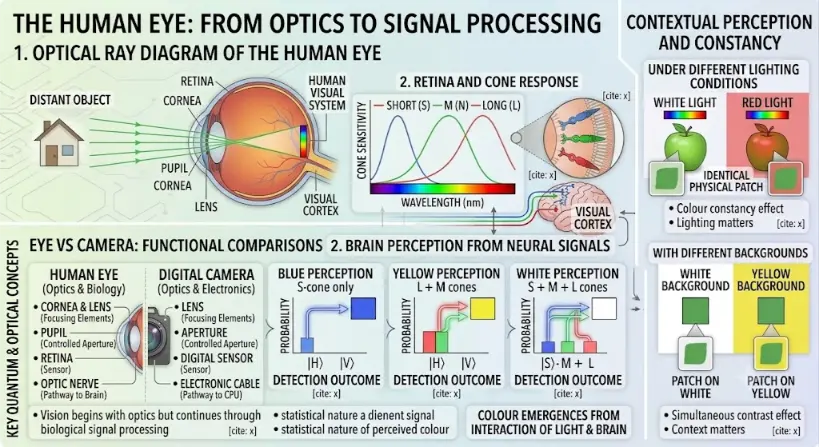
Part 1: Optical Ray Diagram of the Human Eye
The top-left panel outlines the classical geometrical optics of vision:- Light Source: On the far left, an icon of a house represents a “DISTANT OBJECT.”
- Ray Tracing: Parallel green light rays extend from the object toward a cross-section of the human eye. The rays physically bend as they pass through the curved outer Cornea, enter through the opening of the Pupil, and are focused by the internal crystalline Lens.
- Image Formation: The converging rays cross inside the vitreous body and project a crisp, inverted (upside-down), and miniature multicolored image of the house directly onto the Retina—the light-sensitive layer spanning the back wall of the eye.
Part 2: Eye vs. Camera Functional Comparisons
The bottom-left panel features a structured, direct technical comparison detailing how biological organs match modern electronics:| HUMAN EYE (Optics & Biology) | DIGITAL CAMERA (Optics & Electronics) |
|---|---|
| Cornea & Lens: Function as the primary biological Focusing Elements. | Lens Assembly: Functions as the physical Focusing Elements. |
| Pupil / Iris: Acts as a variable, Controlled Aperture adjusting light intake. | Mechanical Aperture: Acts as a variable, Controlled Aperture adjusting light intake. |
| Retina: Acts as the biological light Sensor capturing the image plane. | Digital Sensor: Acts as the electronic image Sensor capturing the image plane. |
| Optic Nerve: Functions as the Pathway to the Brain. | Electronic Cable: Functions as the digital Pathway to the CPU. |
Part 3: Retina Response and Brain Perception
The middle panels illustrate how those focused rays are converted into perceptual data:- Retinal Cones: A circular breakout diagram displays the microscopic Short (S), Medium (M), and Long (L) cone photodetectors inside the retina tissue. An adjacent chart plots three overlapping cone sensitivity curves across the visible wavelength spectrum (measured in nanometers).
- Neural Signal Paths: Colored nerve lines travel from the back of the eye toward a miniature graphic of a human brain, targeting the labeled Visual Cortex.
- Perception Histograms: The lower central zone displays three distinct data blocks mapping out how the brain decrypts color from neural streams. Separate probability histograms chart “Blue Perception” (from S-cones only), “Yellow Perception” (from L + M cones), and “White Perception” (from a combination of S + M + L cones). A text label affirms: “Colour emerges from the interaction of light and the brain.”
Part 4: Contextual Perception and Constancy
A distinct vertical sidebar running down the right-hand side demonstrates the brain’s advanced adjustments over raw optical data:- Lighting Variations: Visualizes a green apple placed under uniform “White Light” versus uniform “Red Light.” Isolated color patches highlight that while the physical wavelengths bouncing off the surface change drastically due to lighting, the brain leverages a “Colour constancy effect” to stabilize perception.
- Background Variations: Shows two identical green square patches placed onto contrasting “White” and “Yellow” backgrounds. Zoom callouts track how a “Simultaneous contrast effect” causes the identical physical square to visually shift and appear different to the viewer depending entirely on its contextual surroundings.
The Path of Light Through the Eye
| Step | Eye Structure | Optical Role |
|---|---|---|
| 1 | Cornea | First major refracting surface; bends incoming light strongly |
| 2 | Aqueous humour | Transparent fluid through which light travels after the cornea |
| 3 | Pupil | Opening that controls how much light enters the eye |
| 4 | Lens | Adjustable focusing element that helps form a sharp image |
| 5 | Vitreous humour | Transparent gel-like medium through which light travels toward the retina |
| 6 | Retina | Light-sensitive neural tissue where the optical image is detected |
| 7 | Optic nerve | Carries visual information toward the brain |
The Cornea: The Main Focusing Surface
The Iris and Pupil: The Eye’s Aperture System
| Pupil Condition | Optical Effect | Everyday Meaning |
|---|---|---|
| Small pupil | Less light enters; depth of focus may improve | Useful in bright environments |
| Large pupil | More light enters; aberrations may become more noticeable | Useful in dim environments |
| Rapid pupil change | Adjusts light entry dynamically | Helps vision adapt between bright and dark scenes |
The Lens: Adjustable Focusing
Accommodation: Changing Focus for Near and Far Objects
| Viewing Situation | Lens Shape | Optical Power | Purpose |
|---|---|---|---|
| Distant object | Flatter lens | Lower power | Focus distant rays on the retina |
| Near object | Rounder lens | Higher power | Focus diverging near rays on the retina |
The Retina: Where the Image Becomes a Signal
| Photoreceptor Type | Main Function | Student-Friendly Meaning |
|---|---|---|
| Rods | Detect low light levels | Important for dim-light and night vision |
| Cones | Detect colour and fine detail | Important for daylight vision and detailed central vision |
The Fovea: The Centre of Sharp Vision
Image Formation on the Retina
The Eye as a Thin-Lens Approximation
Lens Power and Diopters
| Focal Length | Lens Power | Meaning |
|---|---|---|
| 1.0 m | 1 D | Weak focusing power |
| 0.5 m | 2 D | Stronger focusing power |
| 0.25 m | 4 D | Even stronger focusing power |
The Eye and Refractive Errors
| Condition | Basic Optical Problem | Simple Description |
|---|---|---|
| Myopia | Distant light focuses in front of the retina | Distant objects appear blurred |
| Hyperopia | Light tends to focus behind the retina | Near vision may be difficult, depending on age and accommodation |
| Astigmatism | Curvature differs in different directions | Images may blur or distort along certain orientations |
| Presbyopia | Lens loses flexibility with age | Near focusing becomes harder |
Depth of Focus and the Pupil
Optical Aberrations in the Eye
| Aberration Type | Simple Meaning | Possible Visual Effect |
|---|---|---|
| Spherical aberration | Peripheral rays and central rays do not focus at exactly the same point | Reduced sharpness or contrast |
| Chromatic aberration | Different wavelengths focus slightly differently | Small colour-dependent focus effects |
| Astigmatic aberration | Focusing power differs across directions | Line-like blur or directional distortion |
| Higher-order aberrations | More complex imperfections in the wavefront | Glare, halos, or reduced optical quality in some conditions |
The Eye Compared with a Camera
| Camera Component | Eye Component | Similar Role |
|---|---|---|
| Camera lens | Cornea and lens | Focus light |
| Aperture | Pupil | Controls light entry |
| Image sensor | Retina | Detects the image |
| Autofocus system | Accommodation and eye movements | Adjusts focus and attention |
| Image processor | Retina and brain | Interprets visual information |
Resolution of the Eye
Colour and Light Sensitivity
Eye Movements and Visual Attention
Applications of Eye Optics
Vision Correction
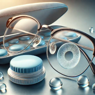
Glasses, contact lenses, and intraocular lenses correct refractive errors by changing how light focuses before it reaches the retina.
Ophthalmic Instruments
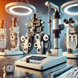
Tools such as slit lamps, fundus cameras, autorefractors, wavefront sensors, and OCT systems depend on eye optics.
Retinal Imaging
Imaging systems use the eye’s optical pathway to observe the retina, blood vessels, optic nerve region, and retinal layers.
Visual Ergonomics
Screen distance, contrast, brightness, glare control, text rendering, and font size affect visual comfort and performance.
Display Design
Displays must account for eye resolution, colour perception, contrast sensitivity, brightness, refresh rate, and viewing distance.
Augmented Reality
AR devices must project images into the user’s visual system while managing focus, field of view, eye tracking, and comfort.
Vision Science
Researchers study how optics, retina, brain, movement, and perception combine to create visual experience.
Biomedical Engineering
Eye optics informs medical devices, diagnostic instruments, corrective lenses, imaging systems, and vision-assistive technologies.
Connections with Wider Physics and Technology
Light and Optics
The eye uses refraction, focusing, image formation, aperture control, and light detection to support vision.
Geometrical Optics
Ray diagrams, lenses, focal length, real images, and refractive errors are central to modelling the eye.
Wave Optics
Diffraction, interference, resolution, and aberrations influence the limits of visual sharpness.
Bio-Optics
The eye is a living optical system, making it a natural bridge between optics, biology, imaging, and diagnostics.
Optical Coherence Tomography
OCT uses light and interference to image tissue layers, especially in the eye and retina.
Medical Imaging
Eye imaging belongs to the wider family of medical imaging technologies used to observe structure and support clinical assessment.
Artificial Intelligence
AI can support retinal image analysis, screening workflows, eye tracking, and visual-interface technologies when properly validated.
Human-Computer Interaction and UX
Visual comfort, readability, attention, contrast, and display design depend on how the human eye and brain process visual information.
Learning Pathway Within Visual Optics
The Eye as an Optical System
Current page. Learn how the cornea, pupil, lens, retina, and optic nerve work together to form images and begin visual perception.
Vision Correction with Lenses
Understand how glasses, contact lenses, and lens power correct myopia, hyperopia, astigmatism, and presbyopia.
Colour Vision and Visual Perception
Explore how cones, wavelengths, colour mixing, contrast, adaptation, and the brain shape human colour vision and perception.
Displays, AR, and Human Vision
Discover how screens, pixels, refresh rates, contrast, AR optics, eye comfort, and human vision shape modern display design.
Quick Check: The Eye as an Optical System
Quick Check: Eye Optics
Common Misunderstandings About Eye Optics
Misunderstanding 1: The Lens Does All the Focusing
Misunderstanding 2: The Eye Works Exactly Like a Camera
Misunderstanding 3: A Bigger Pupil Always Means Better Vision
Misunderstanding 4: Clear Vision Depends Only on the Eye’s Lens
Misunderstanding 5: The Retinal Image Alone Is What We See
Key Terms
- Aberration
- An imperfection in image formation caused by real optical surfaces and materials.
- Accommodation
- The process by which the lens changes shape to focus objects at different distances.
- Cone cells
- Photoreceptors important for colour vision and fine detail in brighter light.
- Cornea
- The clear front surface of the eye that bends incoming light and provides much of the eye’s focusing power.
- Diopter
- A unit of optical power equal to the reciprocal of focal length in metres.
- Focal length
- The distance from an optical element to the point where parallel rays are focused.
- Fovea
- The central retinal region responsible for the sharpest detailed vision.
- Iris
- The coloured part of the eye that controls the size of the pupil.
- Lens
- The adjustable transparent structure that fine-tunes focus for near and distant objects.
- Optic nerve
- The nerve pathway that carries visual information from the retina toward the brain.
- Pupil
- The opening through which light enters the eye.
- Refractive error
- An optical condition in which light does not focus sharply on the retina without correction.
- Retina
- The light-sensitive neural tissue at the back of the eye where the optical image is detected.
- Rod cells
- Photoreceptors important for dim-light vision.
Review Questions
- What does it mean to describe the eye as an optical system?Answer: It means the eye can be studied as a set of components that control light, including refracting surfaces, an aperture, focusing elements, transparent media, and a light-sensitive detector.
- What is the main optical role of the cornea?Answer: The cornea strongly refracts incoming light and provides much of the eye’s focusing power.
- What is the function of the pupil?Answer: The pupil controls the amount of light entering the eye by changing size under the control of the iris.
- What does the lens do?Answer: The lens fine-tunes focus by changing shape, allowing the eye to focus on objects at different distances.
- What is accommodation?Answer: Accommodation is the process by which the eye changes lens shape and optical power to focus near or distant objects on the retina.
- Why is the retina important?Answer: The retina detects the focused optical image and begins converting light patterns into neural signals.
- What is the fovea?Answer: The fovea is the central retinal region responsible for the sharpest detailed vision.
- Why is the eye-camera comparison useful but incomplete?Answer: Both systems focus light onto a detecting surface, but the eye is living neural tissue connected to active brain processing, movement, and perception.
Thought-Provoking Questions
- Why is vision both a physics problem and a biology problem?Answer: Physics explains how light is focused on the retina, while biology explains how retinal cells and the brain convert that light pattern into visual experience.
- Why might a perfectly sharp retinal image still not guarantee perfect visual perception?Answer: Perception also depends on retinal health, optic nerve function, brain processing, attention, contrast, adaptation, and visual context.
- How does accommodation show that the eye is an active optical system?Answer: The eye changes its optical power to focus at different distances, rather than behaving like a fixed lens.
- Why does display design need to understand eye optics?Answer: Displays must consider resolution, viewing distance, brightness, contrast, colour, focus, glare, and visual comfort.
- How might future AR systems become more comfortable?Answer: They may improve by reducing latency, improving tracking, managing depth cues, increasing optical quality, using eye tracking, and addressing vergence-accommodation conflict.
Comprehensive Numerical Problems with Solutions
- A simplified eye model uses a converging lens. An object is placed 25 cm from the lens and a real image forms 2 cm behind the lens. Find the focal length.Solution:Apply the thin lens coordinate formula:$$\frac{1}{f} = \frac{1}{d_o} + \frac{1}{d_i}$$Substitute \(d_o = 25\,\text{cm}\) and \(d_i = 2\,\text{cm}\):$$\frac{1}{f} = \frac{1}{25} + \frac{1}{2} = 0.04 + 0.50 = 0.54\,\text{cm}^{-1}$$$$f = \frac{1}{0.54} \approx 1.8518\,\text{cm}$$Answer: The effective focal length is approximately 1.85 cm.
- A person with hyperopia has a near point of 50 cm. What lens power is required to see objects clearly at a comfortable distance of 25 cm?Solution:1. The corrective lens must redirect light originating from an object at \(d_o = 25\,\text{cm}\) so that it appears to diverge from the patient’s natural near point as a virtual image (\(d_i = -50\,\text{cm}\)).2. Compute reciprocal focal tracking lengths:$$\frac{1}{f} = \frac{1}{25} – \frac{1}{50} = \frac{2}{50} – \frac{1}{50} = \frac{1}{50}\,\text{cm}^{-1}$$$$f = 50\,\text{cm} = 0.50\,\text{m}$$3. Extract optical refractive power parameters in diopters:$$P = \frac{1}{f_{\text{meters}}} = \frac{1}{0.50\,\text{m}} = +2.0\,\text{D}$$Answer: A +2.0 D converging lens is required.
- A corrective lens for a myopic person has a focal length of −5 cm. If a target tracking object is positioned 25 cm away, find the calculated image distance.Solution:Use the thin lens equation:$$\frac{1}{f} = \frac{1}{d_o} + \frac{1}{d_i}$$Substitute \(f = -5\,\text{cm}\) and \(d_o = 25\,\text{cm}\):$$\frac{1}{-5} = \frac{1}{25} + \frac{1}{d_i}$$$$-0.20 = 0.04 + \frac{1}{d_i}$$$$\frac{1}{d_i} = -0.20 – 0.04 = -0.24\,\text{cm}^{-1}$$$$d_i = \frac{1}{-0.24} \approx -4.1667\,\text{cm}$$Answer: The image is virtual and forms precisely 4.17 cm in front of the lens matrix (on the same side as the object).
- A clinical model dictates that an eye has an effective structural focal length of 1.8 cm. Find its absolute optical power.Solution:1. Scale the focal parameters from centimeters to standard meters:$$1.8\,\text{cm} = 0.018\,\text{m}$$2. Take the reciprocal quotient to isolate diopters:$$P = \frac{1}{f} = \frac{1}{0.018\,\text{m}} \approx 55.5556\,\text{D}$$Answer: The absolute optical baseline power measures approximately 55.6 D.
- A person has an uncorrected far point of 100 cm. What lens power is needed to establish comfortable distant vision at infinity?Solution:For parallel rays from infinity to focus properly, the corrective lens must project incoming light back to form a virtual image at the patient’s far point. The lens focal length must equal \(f = -100\,\text{cm} = -1.0\,\text{m}\).$$P = \frac{1}{f} = \frac{1}{-1.0\,\text{m}} = -1.0\,\text{D}$$Answer: A −1.0 D diverging lens is required.
- An iris contracts so that the pupil aperture diameter increases from 2 mm up to 6 mm. By what factor does the total light collection area increase?Solution:Ocular aperture area scales proportionally to the square of the diameter parameter (\(A \propto d^2\)):$$\text{Area Factor} = \left(\frac{d_{\text{final}}}{d_{\text{initial}}}\right)^2 = \left(\frac{6\,\text{mm}}{2\,\text{mm}}\right)^2 = (3)^2 = 9$$Answer: The complete pupil collection area increases by a factor of 9.
- A display font stroke features a physical height parameter of 3 mm and is viewed from a distance coordinate of 0.60 m. Estimate its angular height in radians using the small-angle approximation.Solution:1. First convert the text stroke dimension to uniform meters:$$3\,\text{mm} = 0.003\,\text{m}$$2. Apply the arc-length approximation ratio (\(\theta \approx \frac{s}{d}\)):$$\theta \approx \frac{0.003\,\text{m}}{0.60\,\text{m}} = 0.005\,\text{rad}$$Answer: The target angular height measures exactly 0.005 radians.
- Two thin converging ophthalmic lenses are placed in direct contact, featuring separate focal lengths of 10 cm and 15 cm. Find the effective focal length of the combined array.Solution:The cumulative power of lenses in contact equals the sum of their individual diopters:$$\frac{1}{f_{\text{eff}}} = \frac{1}{f_1} + \frac{1}{f_2} = \frac{1}{10} + \frac{1}{15} = \frac{3}{30} + \frac{2}{30} = \frac{5}{30} = \frac{1}{6}\,\text{cm}^{-1}$$$$f_{\text{eff}} = 6\,\text{cm}$$Answer: The net effective focal length of the combined system is exactly 6 cm.
External References for Further Reading
- National Eye Institute: How the Eyes Work — A clear explanation of the cornea, pupil, lens, retina, and optic nerve.
- OpenStax College Physics: Physics of the Eye — A student-friendly physics explanation of the eye, focusing, and image formation.
- NCBI Bookshelf: The Formation of Images on the Retina — A useful neuroscience reference on how the cornea and lens focus images on the retina.
- NCBI Bookshelf: Anatomy of the Retina — A reference on retinal structure and the role of photoreceptors.
- American Academy of Ophthalmology: Eye Anatomy — A public-facing guide to the main parts of the eye and their roles.
- OpenStax University Physics: The Eye — A more advanced physics section on the eye and optical focusing.