
Protein synthesis is one of the most fundamental biological processes that sustains life, converting genetic information into functional proteins. This intricate process, central to the fields of science and biology, unfolds in two major stages: transcription and translation. It begins with the instructions encoded in DNA, studied in DNA and RNA, and results in chains of amino acids forming proteins with diverse functions in the body. Understanding this mechanism is pivotal for anyone delving into genetics or molecular genetics.
To grasp the complexity of protein synthesis, one must appreciate the roles of cellular structures covered in cell biology, including cell structure and cell physiology. The ribosomes, endoplasmic reticulum, and other organelles orchestrate the translation of mRNA into polypeptides, while cell communication mechanisms regulate gene activity to meet developmental and environmental demands. This regulation is essential in processes like the cell cycle and cell development.
The accurate transmission of genetic information during molecular basis of inheritance is crucial to ensuring that protein synthesis occurs correctly. When mutations arise, as explored in genetic mutation, they may alter protein function, with implications ranging from benign variations to serious genetic disorders. Advances in DNA technology and molecular techniques in research allow scientists to manipulate genes and study protein synthesis in unprecedented detail.
Explorations in gene expression reveal how not all genes are active at all times, but are turned on or off in specific cells and conditions. This selective activation is crucial for differentiation and physiological function. The broader implications of these mechanisms extend to fields like genomics and population genetics, which examine genetic variation within and across populations. Quantitative perspectives in quantitative genetics allow for statistical analysis of traits governed by multiple genes involved in protein production.
Protein synthesis also connects with evolutionary dynamics, as evidenced in molecular evolution and evolutionary biology. Mutations and selective pressures shape the coding sequences that determine protein structure and function. Researchers further apply insights from Mendelian genetics and applications of genetics in medicines to diagnose and treat genetic diseases. Ultimately, studying protein synthesis opens doors to biotechnology, synthetic biology, and medical advances that continue to reshape our understanding of life at the molecular level.
Whether exploring the origin of proteins in unicellular organisms in ecology, or understanding systemic regulation at a multicellular level, protein synthesis remains a cornerstone of biological inquiry. It is where the language of DNA becomes the machinery of life—a transformative process central to all living systems.
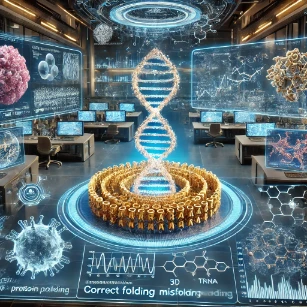
From Code to Function: Translating Genetic Information into Proteins
Molecular Genetics – Overview
Connects genetic coding with functional biological molecules that drive essential cellular processes and cellular metabolism.
Protein Synthesis
Describes the coordinated processes of transcription and translation that convert linear genetic sequences into functional proteins.
DNA & RNA
Provides the master nuclear templates and messenger intermediate molecules required for polypeptide production.
Gene Expression
Regulates when and how specific proteins are synthesized in response to physiological and developmental cues.
Genetic Mutation
Alters polypeptide primary structures and folding stability by changing underlying nucleotide coding sequences.
Inheritance
Transmits protein-coding alleles that determine structural, enzymatic, and physiological phenotypic traits across generations.
Molecular Evolution
Examines how subtle amino acid sequence variations accumulate across evolutionary timescales to drive adaptive innovation.
DNA Technology
Engineers expression vectors and recombinant systems to manufacture custom proteins for research and therapeutic applications.
Molecular Techniques
Employs specialized assays such as Western blotting and ribo-seq to measure translational efficiency and protein levels.
Applications in Medicine
Develops targeted pharmacological agents, antibiotics, and biologics that specifically modulate protein translation machinery.
The Genetic Code and How It Translates into Proteins
The genetic code is the universal chemical dictionary that defines how nucleotide triplets in mRNA are translated into specific amino acid sequences, constructing functional polypeptide chains.
Properties of the Genetic Code
The genetic code exhibits four major structural characteristics:
1. Universal Nature: With minor exceptions in mitochondrial genomes and specific protozoans, the same codon assignments are utilized by bacteria, plants, animals, and viruses.
2. Degeneracy / Redundancy: With 64 possible triplet combinations (43) coding for 20 standard amino acids and 3 stop signals, multiple distinct codons specify the same amino acid. For instance, leucine, arginine, and serine are each coded by six different mRNA codons.
3. Non-Overlapping Reading Frames: During translation, the ribosome reads nucleotides in contiguous, discrete triplets without sharing nucleotides between adjacent codons.
4. Triplet Codons: Each codon consists of three consecutive RNA nucleotides. The universal start codon is 5′-AUG-3′, which codes for methionine (N-formylmethionine in prokaryotes) and establishes the correct reading frame. The three stop codons—UAA (ochre), UAG (amber), and UGA (opal)—do not code for amino acids; instead, they bind release factors to terminate translation.
Core Stages of Protein Synthesis
Protein synthesis takes place in two tightly linked sequential stages:
1. Transcription (DNA → pre-mRNA → mRNA): In eukaryotic nuclei, RNA polymerase II transcribes a gene’s template DNA strand into a pre-mRNA precursor. Processing steps—including 5′ capping, 3′ polyadenylation, and intron splicing via the spliceosome—generate mature mRNA, which is exported to the cytoplasm.
2. Translation (mRNA → Polypeptide): Cytoplasmic translation executes in three distinct sub-phases:
• Initiation: The small ribosomal subunit (40S in eukaryotes) binds the 5′ cap of mRNA and scans for the AUG start codon. An initiator tRNA carrying methionine (tRNAiMet) pairs its 3′-UAC-5′ anticodon with AUG. The large ribosomal subunit (60S) then joins, forming a functional 80S ribosome with the initiator tRNA positioned in the peptidyl (P) site.
• Elongation: An aminoacyl-tRNA complementary to the second codon enters the aminoacyl (A) site assisted by elongation factors (EF-Tu / eEF1A). Peptidyl transferase, a ribozyme component of the large subunit rRNA, catalyzes peptide bond formation between the P-site amino acid and the A-site amino acid. Translocation driven by EF-G / eEF2 shifts the ribosome three nucleotides forward, moving the deacylated tRNA to the exit (E) site for discharge, while the growing peptidyl-tRNA moves from A to P site.
• Termination: When an A-site codon encounters UAA, UAG, or UGA, eukaryotic release factors (eRF1 and eRF3) enter the A site, cleaving the ester bond holding the polypeptide to the P-site tRNA. The completed polypeptide is liberated, and the ribosomal complex dissociates into free subunits.
Translation Machinery Components
Translation relies on three core macromolecular structures:
• Messenger RNA (mRNA): The single-stranded transcript containing coding sequences flanked by 5′ and 3′ untranslated regions (UTRs).
• Transfer RNA (tRNA): Cloverleaf-shaped adaptor RNAs containing a specific 3-base anticodon loop at one end and a 3′-CCA amino acid attachment site at the other, charged by aminoacyl-tRNA synthetases.
• Ribosomes: Complex ribonucleoprotein machines. Eukaryotic 80S ribosomes consist of a 40S small subunit (18S rRNA + 33 proteins) responsible for decoding and a 60S large subunit (28S, 5.8S, 5S rRNAs + 47 proteins) responsible for catalytic peptide bond synthesis.
Post-Translational Modifications and Their Importance
Following ribosomal synthesis, nascent polypeptide chains undergo enzymatic covalent adjustments termed post-translational modifications (PTMs). PTMs dramatically expand proteomic diversity, regulating enzyme activity, intracellular trafficking, protein stability, and macromolecular interactions.
Major Types of Post-Translational Modifications
1. Phosphorylation: Reversible addition of a phosphate group (ΔPO42-) to the hydroxyl group of serine, threonine, or tyrosine residues catalyzed by protein kinases and reversed by phosphatases. Phosphorylation induces conformational shifts that act as molecular on/off switches; for example, Cyclin-Dependent Kinases (CDKs) require specific phosphorylation events to drive cell cycle transitions.
2. Glycosylation: Enzymatic attachment of oligosaccharide chains to asparagine side chains (N-linked in the ER) or serine/threonine hydroxyl groups (O-linked in the Golgi). Glycosylation governs proper glycoprotein folding, protects against proteolytic degradation, and mediates cell-cell recognition.
3. Acetylation: Transfer of an acetyl group (ΔCOCH3) from acetyl-CoA to lysine residues or the N-terminus of proteins. Histone acetyltransferases (HATs) neutralize positive charges on histone tail lysines, loosening chromatin structure to activate gene transcription.
4. Ubiquitination: Covalent attachment of the 76-amino-acid regulatory protein ubiquitin to target lysine residues via a three-enzyme cascade (E1 activation, E2 conjugation, E3 ligase recognition). Polyubiquitin chains linked via Lysine-48 target damaged or misfolded proteins to the 26S proteasome for degradation.
5. Proteolytic Cleavage: Irreversible peptide bond hydrolysis that converts inactive zymogens or pro-proteins into active mature forms. For example, preproinsulin undergoes signal peptide cleavage in the ER to form proinsulin, followed by endopeptidase cleavage in secretory vesicles to remove C-peptide and yield functional double-chain insulin.
Protein Folding and Its Importance
Protein folding is the physical process by which an unbranched polypeptide chain folds into its thermodynamically stable, biologically active three-dimensional tertiary structure.
Hierarchical Levels of Protein Structure
Protein architecture is organized across four distinct structural tiers:
• Primary Structure: The linear, genetically determined sequence of amino acids linked by covalent peptide bonds.
• Secondary Structure: Highly regular local conformations—predominantly α-helices and β-pleated sheets—stabilized by hydrogen bonding between peptide backbone carbonyl oxygen atoms and amide nitrogen protons.
• Tertiary Structure: The overall 3D spatial fold of a single polypeptide chain, stabilized by side-chain (R-group) interactions including hydrophobic collapse, ionic salt bridges, hydrogen bonding, and covalent disulfide bonds (ΔSΔSΔ) between cysteine residues.
• Quaternary Structure: The spatial arrangement and functional subunit assembly of multimeric proteins comprising two or more polypeptide chains (e.g., the tetrameric α2β2 structure of hemoglobin).
Chaperones and Assisted Folding
Although primary sequences contain all information required for folding (Anfinsen’s dogma), high cytoplasmic protein concentrations induce hydrophobic aggregation. Molecular chaperones—such as Heat Shock Proteins (Hsp70, Hsp90) and chaperonins (GroEL/GroES in bacteria, TRiC in eukaryotes)—bind exposed hydrophobic patches on nascent chains, utilizing ATP hydrolysis to prevent misfolding and isolate proteins within micro-environments until correct tertiary structures form.
Misfolding of Proteins and Human Diseases
When cellular quality control mechanisms fail, misfolded proteins expose hydrophobic surfaces, forming insoluble β-sheet-rich toxic aggregates and amyloid fibrils that disrupt cellular homeostasis.
Causes of Protein Misfolding
Protein misfolding arises from several intrinsic and extrinsic factors:
• Single nucleotide mutations altering primary amino acid residue chemistry.
• Environmental or physiological stressors (heat shock, oxidative stress, pH shifts).
• Age-related decline in proteostasial capacity, chaperone efficiency, or proteasomal clearance pathways.
Pathology of Protein Misfolding Diseases
Misfolding and aggregation underly several severe chronic human pathologies:
1. Alzheimer’s Disease: Proteolytic cleavage of Amyloid Precursor Protein (APP) by β- and γ-secretases yields β-amyloid (Aβ42) peptides that aggregate into extracellular senile plaques. Concurrently, hyperphosphorylated tau protein forms intracellular neurofibrillary tangles, causing widespread synaptic loss and neurodegeneration.
2. Parkinson’s Disease: Misfolded α-synuclein oligomerizes into insoluble fibrillar inclusions termed Lewy bodies within dopaminergic neurons of the substantia nigra, leading to motor tremors and progressive akinesia.
3. Cystic Fibrosis: The common ΔF508 deletion in the CFTR gene causes misfolding of the CFTR chloride channel in the endoplasmic reticulum. ER quality control degrades the misfolded protein via ER-associated degradation (ERAD), preventing its delivery to the plasma membrane and causing thick mucus accumulation in pulmonary passages.
4. Huntington’s Disease: An unstable expansion of CAG trinucleotide repeats in the HTT gene encodes an elongated polyglutamine (polyQ) tract in the huntingtin protein, driving toxic intracellular nuclear inclusions in striatal neurons.
5. Prion Diseases (Transmissible Spongiform Encephalopathies): Infectious prion proteins (PrPSc) rich in β-sheets bind endogenous alpha-helical cellular prion proteins (PrPC), autocatalytically inducing them to misfold into toxic aggregates. Examples include Creutzfeldt-Jakob Disease (CJD) in humans and Bovine Spongiform Encephalopathy (BSE).
Summary of Core Protein Synthesis Concepts
| Process / Topic | Key Molecular Mechanisms & Functions |
|---|---|
| Genetic Code | Universal, degenerate triplet mRNA codons that specify amino acids or signal start/stop translation. |
| Translation | Ribosome-catalyzed decoding of mRNA into polypeptides via initiation, elongation, and termination phases. |
| Post-Translational Modifications | Enzymatic alterations (phosphorylation, glycosylation, cleavage) that modulate activity, localization, and degradation. |
| Protein Folding | Hierarchical folding into functional 3D structures assisted by molecular chaperones to prevent aggregation. |
| Protein Misfolding Diseases | Accumulation of toxic protein aggregates causing neurodegenerative disorders (Alzheimer’s, Parkinson’s, Prions). |
Why Study Protein Synthesis
Central Role in Gene Expression
Protein synthesis is the process by which cells produce proteins based on genetic instructions. It involves transcription of DNA into RNA and translation of RNA into proteins. Understanding this process is key to learning how cells function. It connects genetics to cellular and physiological activity.
Understanding Ribosomes and Translation
Students learn how ribosomes assemble amino acids into polypeptide chains using mRNA templates. They explore the roles of tRNA, rRNA, and initiation factors in translation. This provides insight into the mechanics of protein production. It enhances comprehension of cellular operations and molecular biology.
Relevance to Health and Disease
Errors in protein synthesis can lead to genetic disorders, cancer, and neurodegenerative diseases. Studying this process enables students to understand the molecular basis of these conditions. This knowledge is crucial for developing medical treatments and diagnostic tools. It supports careers in healthcare and biomedical research.
Biotechnological Applications
Protein synthesis is used in the production of insulin, enzymes, and therapeutic proteins. Students learn how to express and purify proteins in bacterial or mammalian systems. These skills are vital in pharmaceutical and biotech industries. It enables students to participate in developing biological products.
Integration with Molecular Genetics
Studying protein synthesis integrates knowledge of genes, DNA structure, and molecular machinery. It helps students see how genetic information is translated into cellular function. This comprehensive understanding prepares them for research in molecular and cellular biology. It also strengthens their foundation in life sciences.
About This Interactive Simulator:
This interactive simulator models the kinetic dynamics of mRNA translation during protein synthesis. Adjust the mRNA transcript length, elongation rate, and ribosome initiation interval to compute overall translation runtime, ribosome density per mRNA (polysome traffic), and polypeptide production rates.
- mRNA Coding Length (Codons): Set transcript length (100 to 2,000 codons).
- Elongation Rate (aa/sec): Adjust translation speed (2 to 20 amino acids per second).
- Initiation Interval (sec): Control how frequently a new ribosome engages the AUG start codon (1 to 10 seconds).
Interactive Translation Kinetics & Polysome Simulator
Applications of Protein Synthesis in Medicine, Biotechnology, and Research
Understanding protein synthesis machinery has yielded major clinical, pharmaceutical, and biotechnological innovations. Because cellular protein production is strictly controlled, therapeutic interventions can selectively target translational components in pathogens or diseased tissues.
In pharmacology, major antibiotic classes exploit structural differences between prokaryotic 70S ribosomes and eukaryotic 80S ribosomes. Aminoglycosides (e.g., streptomycin) bind the 30S bacterial subunit to induce mRNA misreading, tetracyclines block aminoacyl-tRNA entry into the A site, and macrolides (e.g., erythromycin) bind the 50S subunit to inhibit translocation along the mRNA.
In biotechnology, recombinant protein manufacturing utilizes optimized plasmid expression vectors in E. coli or Chinese Hamster Ovary (CHO) cells to produce therapeutic proteins, including human insulin, growth hormone, clotting factors, and monoclonal antibodies. Furthermore, synthetic mRNA therapeutics (e.g., mRNA vaccines) deliver lipid-nanoparticle-encapsulated transcripts directly into human cells, instructing host ribosomes to translate immunogenic viral proteins (such as viral spike proteins) and elicit protective immune responses.
Emerging Trends, Challenges & Future Directions
Research in protein synthesis is rapidly expanding into single-molecule translation tracking, cell-free synthetic biology, and bio-orthogonal protein engineering. Ribosome profiling (Ribo-Seq) now enables global genome-wide mapping of active translation at single-codon resolution by sequencing ribosome-protected mRNA fragments.
Cell-free protein synthesis (CFPS) systems bypass living cell membranes, allowing rapid in vitro production of toxic or complex membrane proteins. Concurrently, expanded genetic code technology incorporates unnatural synthetic amino acids (uAAs) into target proteins using engineered orthogonal aminoacyl-tRNA synthetase / tRNA pairs that respond to amber stop codons, enabling novel biophysical probes and site-specific drug conjugations.
Key challenges include resolving structural details of translational stalling on complex non-coding structures, mitigating off-target immune responses to synthetic RNA therapies, and engineering targeted protein degradation platforms (PROTACs) to clear pathogenic protein aggregates in neurodegenerative conditions.
Frequently Asked Questions
What is protein synthesis?
Protein synthesis is the process by which cells build proteins based on genetic instructions encoded in DNA. It involves transcription of DNA into RNA and translation of RNA into a specific sequence of amino acids that fold into a functional protein.
What are the two main stages of protein synthesis?
The two main stages are transcription and translation. In transcription, a segment of DNA is copied into messenger RNA (mRNA). In translation, the mRNA code is read by ribosomes and transfer RNAs (tRNAs) to assemble amino acids in the correct order to form a protein.
How does transcription work?
During transcription, an enzyme called RNA polymerase binds to a gene’s promoter region, separates the DNA strands, and synthesizes a complementary RNA strand based on the DNA template. The resulting pre-mRNA may be processed (splicing, 5′ cap, poly-A tail) before becoming mature mRNA that travels out of the nucleus to the ribosome.
How does translation work?
During translation, ribosomes read the sequence of codons on the mRNA. Transfer RNAs (tRNAs), each carrying a specific amino acid, match their anticodon to each codon on the mRNA. The ribosome links the amino acids together, forming a polypeptide chain that folds into a functional protein.
What roles do ribosomes, mRNA, and tRNA play in protein synthesis?
Ribosomes are molecular machines where proteins are assembled. mRNA carries the genetic code from DNA to the ribosome. tRNA brings amino acids to the ribosome and matches them to the mRNA codons, ensuring the correct sequence of amino acids in the protein.
Why is protein folding and post-translational modification important?
Protein folding ensures the newly formed polypeptide folds into the correct three-dimensional shape required for its function. Post-translational modifications — such as phosphorylation, glycosylation, or cleavage — further modify proteins to activate them, direct them to specific cellular locations, or regulate their stability and activity.
How can errors in protein synthesis affect organisms?
Errors during transcription, translation, folding, or post-translational modification can produce malfunctioning or misfolded proteins, which may disrupt normal cellular processes. Such errors can lead to diseases, metabolic disorders, or developmental problems if key proteins fail to function correctly.
Why is understanding protein synthesis important for further study and careers?
Understanding protein synthesis is fundamental for studies in molecular biology, genetics, biochemistry, and medicine. It underpins how traits are expressed, how cells function, how diseases occur, and how medical treatments such as protein-based drugs are developed. This knowledge is essential for careers in research, healthcare, biotechnology, and pharmaceuticals.
End-of-Page Exercises & Practice Module
Section 1: Foundational Review Questions
Test your baseline knowledge of transcription, translation, and protein folding using these collapsible practice questions:
1. What is the start codon, and what amino acid does it incorporate in eukaryotes?
The universal start codon is 5′-AUG-3′. In eukaryotes, it codes for the amino acid methionine (Met), which forms the N-terminal amino acid of nascent polypeptide chains prior to processing.
2. What are the three stop codons, and how do they terminate translation?
The three stop codons are 5′-UAA-3′, 5′-UAG-3′, and 5′-UGA-3′. They do not bind aminoacyl-tRNAs; instead, they bind release factors (eRF1 in eukaryotes) that trigger ester bond hydrolysis between the peptidyl chain and the P-site tRNA.
3. What catalytic entity forms peptide bonds during translation elongation?
Peptide bond formation is catalyzed by peptidyl transferase, which is a ribozyme component (28S rRNA in eukaryotic 60S large subunits; 23S rRNA in prokaryotic 50S large subunits) of the ribosome.
4. How does phosphorylation alter protein function?
Adding a negatively charged phosphate group (ΔPO42-) to serine, threonine, or tyrosine residues induces local steric and electrostatic shifts, altering enzyme catalytic activity, cellular localization, or protein-protein binding interactions.
5. What role do chaperones play in cell physiology?
Molecular chaperones (e.g., Hsp70, GroEL) prevent hydrophobic aggregation of exposed polypeptide regions during translation or heat stress, promoting correct 3D tertiary folding.
6. How does ubiquitination regulate protein turnover?
Attach Polyubiquitin chains linked via Lysine-48 residue tags misfolded, damaged, or regulatory proteins for recognition and proteolysis inside the 26S proteasome complex.
7. What structural features characterize protein secondary structure?
Secondary structures consist of regular, localized spatial arrangements—primarily α-helices and β-pleated sheets—stabilized by hydrogen bonds along the peptide backbone.
Section 2: Analytical & Scenario-Based Questions
1. Explain wobble hypothesis and its role in genetic code degeneracy.
Crick’s wobble hypothesis states that non-standard base pairing can occur between the 5′ base of the tRNA anticodon and the 3′ base (third position) of the mRNA codon. This wobble flexibility allows a single tRNA species containing modified bases (such as inosine) to pair with multiple synonymous mRNA codons, reducing the minimum required tRNA species from 61 to approximately 31.
2. Why do single nucleotide insertions cause more severe phenotypic damage than single amino acid substitutions?
Single amino acid substitutions (missense mutations) alter a single codon, changing only one amino acid residue in the polypeptide. Single nucleotide insertions shift the non-overlapping reading frame downstream of the mutation (frameshift mutation), completely altering all subsequent downstream amino acid assignments and typically introducing premature stop codons.
3. How do tetracycline antibiotics selectively inhibit bacterial translation without harming human host cells?
Tetracyclines specifically bind to the 30S small subunit of bacterial 70S ribosomes, physically blocking charged aminoacyl-tRNA access to the A site. Eukaryotic cytoplasm utilizes 80S ribosomes containing a 40S small subunit with structural differences that prevent high-affinity tetracycline binding at therapeutic concentrations.
4. Why is extracellular protein accumulation toxic in Alzheimer’s disease?
Self-assembling β-amyloid oligomers expose hydrophobic patches that disrupt neuronal plasma membrane integrity, trigger excessive calcium influx, induce neuroinflammation via microglial activation, and impair synaptic transmission in hippocampal circuits.
5. What is the role of signal peptidase in secretory protein synthesis?
N-terminal signal peptides direct translating ribosome-mRNA complexes to the Rough Endoplasmic Reticulum (RER) membrane. As the polypeptide translocates into the ER lumen, signal peptidase cleaves the hydrophobic signal sequence, releasing the mature precursor for luminal folding.
6. How does ricin toxin inhibit eukaryotic protein synthesis?
Ricin contains an A-chain ribosome-inactivating protein (RIP) that functions as an N-glycosidase. It depurinates a specific adenine residue (A4324) in the 28S rRNA of the 60S subunit, preventing elongation factor binding and halting translation permanently.
7. Contrast Homology-Directed Repair with Non-Homologous End Joining during CRISPR editing.
Non-Homologous End Joining (NHEJ) directly ligates double-stranded breaks without a template, frequently inserting or deleting nucleotides to knock out genes. Homology-Directed Repair (HDR) uses an exogenous homologous donor template to incorporate precise sequence edits or new genes.
Section 3: Step-by-Step Numerical Problems
Problem 1: Polypeptide Length and mRNA Coding Requirement Calculation
A functional eukaryotic protein consists of 450 amino acid residues. Calculate: (a) the minimum number of nucleotides required in the mRNA coding region (including start and stop codons), and (b) the total number of high-energy phosphate bonds (ΔP) consumed to synthesize this polypeptide chain during translation elongation.
Solution:
(a) Each amino acid requires a 3-nucleotide codon. For 450 amino acids:
450 amino acids × 3 nucleotides/codon = 1,350 nucleotides.
Adding 1 stop codon (3 nucleotides):
1,350 + 3 = 1,353 nucleotides.
(b) Energetics of translation:
• Charging each tRNA consumes 2 high-energy phosphate bonds (ATP → AMP + 2 Pi):
450 amino acids × 2 ATP = 900 high-energy bonds.
• Initiation consumes 1 GTP = 1 high-energy bond.
• Elongation consumes 2 GTP per peptide bond (1 for EF-Tu/eEF1A entry, 1 for EF-G/eEF2 translocation):
449 peptide bonds × 2 GTP = 898 high-energy bonds.
• Termination consumes 1 GTP = 1 high-energy bond.
Problem 2: Translation Time Calculation
If a eukaryotic ribosome translocates along mRNA at an average speed of 6 codons per second, how many seconds will it take to synthesize a 360-amino acid protein from initiation to termination?
Solution:
The total coding length is 360 codons.
Using the velocity equation:
Problem 3: Polysome Ribosome Spacing Calculation
An active mRNA transcript is 1,200 nucleotides long in its coding region. If ribosomes on this polyribosome (polysome) are spaced at a minimal physical distance of 80 nucleotides apart, determine the maximum number of ribosomes that can translate this single mRNA simultaneously.
Solution:
Calculate maximum ribosome occupancy:
Problem 4: Protein Molecular Weight Estimation
Estimate the molecular weight (in Daltons and kDa) of a globular protein encoded by an open reading frame (ORF) containing 990 base pairs (excluding the stop codon), assuming the average molecular weight of an amino acid residue is 110 Da.
Solution:
First, determine the number of amino acids encoded:
Number of amino acids = 990 bp / 3 bp/codon = 330 amino acids.
Multiply by average residue weight:
36,300 Da / 1,000 = 36.3 kDa.
Problem 5: Combinatorial Triplet Codon Determination
If a synthetic organism utilized an expanded genetic code with 6 distinct nitrogenous bases (A, T, C, G, X, Y) arranged in triplet codons, how many total unique codons could be constructed?
Solution:
Use the permutation formula for triplet codons with N possible bases:
Total Codons = 63 = 6 × 6 × 6 = 216 unique triplet codons.
Problem 6: Radioisotope Incorporation and Protein Yield
In an in vitro translation assay incorporating 35S-methionine, a protein contains 4 methionine residues per molecule. If 2.0 nanomoles of 35S-methionine are fully incorporated into complete protein products, how many nanomoles of functional protein were synthesized?
Solution:
Each protein molecule incorporates 4 methionine residues:
Problem 7: tRNA Charging Energetic Yield
A cell synthesizes 106 total protein molecules per hour, with an average length of 300 amino acids per protein. Calculate the total ATP energy consumed strictly during the tRNA charging (aminoacylation) phase per hour.
Solution:
Total amino acids incorporated = 106 proteins × 300 aa/protein = 3 × 10⁸ amino acids.
Each tRNA charging reaction converts 1 ATP → AMP + 2 Pi (equivalent to 2 high-energy bonds):