
Orthopedics is the specialty that focuses on bones, joints, muscles, ligaments, and the mechanics of movement. It includes fractures, sports injuries, arthritis, back problems, and many conditions that affect how people walk, lift, work, and live.
Students are often drawn to Orthopedics because it is highly practical. The specialty combines anatomy, imaging, procedures, surgery, and rehabilitation. Many treatments have visible results: less pain, better alignment, stronger movement, or a return to daily activity.
If you are interested in anatomy and enjoy thinking about structure and function together, Orthopedics is a very natural field to explore.
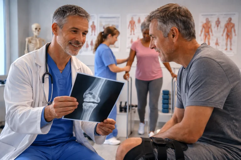
This visual shows an orthopedic doctor explaining a knee X-ray to a patient in a rehabilitation-focused clinic setting, illustrating the practical and structure-based nature of Orthopedics. The scene reflects how this specialty combines anatomy knowledge, imaging, clinical judgment, and recovery planning to treat conditions affecting movement. For students exploring medicine, the image highlights a key strength of Orthopedics: doctors do not only treat injuries or joint problems—they also help patients restore function, reduce pain, and return to normal life. The rehabilitation activity in the background reinforces an important idea in orthopedic care: successful treatment often depends not only on procedures or surgery, but also on careful follow-up and coordinated recovery support.
How Orthopedic Doctors Think About the Body
They focus on structure and movement together
Orthopedic care is not only about whether a bone is broken. It is also about whether the patient can use the limb properly afterward and whether movement can be restored safely.
They use imaging as a core tool
X-rays, CT scans, and MRIs are very important in Orthopedics, but the images must always be matched with the patient’s pain, movement, and physical examination.
They treat both sudden injuries and wear-and-tear
Orthopedics includes trauma care for fractures and acute injuries, but also chronic conditions such as arthritis and spinal degeneration.
They work closely with rehabilitation teams
Even after good treatment or surgery, recovery often depends on physiotherapy and gradual strengthening. Orthopedics and rehabilitation work closely together.
They choose between non-surgical and surgical care
Not every orthopedic problem needs an operation. Doctors must decide whether a cast, brace, exercise program, or procedure will give the best outcome.
Real-Life Cases That Show Orthopedics in Practice
- Example 1: A wrist fracture after a fall. A patient falls and develops swelling and pain around the wrist. The orthopedic doctor reviews the X-ray and decides whether the bone can heal well in a cast or needs surgical fixation.
- Example 2: Knee injury during sport. A young athlete twists the knee and feels a “pop.” Orthopedic assessment may suggest a ligament injury. The doctor examines stability, reviews scans, and plans treatment.
- Example 3: Chronic knee arthritis in an older adult. A patient struggles with walking and stairs because of knee pain. Orthopedics may begin with medications, exercise, and support devices, and later discuss surgery if needed.
- Example 4: Recovery after fracture surgery. After a broken leg is repaired, the orthopedic team continues to monitor healing and function. Orthopedics is not only about “fixing” but also about restoring use of the limb.
A Glimpse into the Daily Work of an Orthopedic Doctor
- Clinic consultations with a strong examination focus: Orthopedic clinic work often involves pain assessment, joint movement testing, and comparison of function before and after treatment.
- Reviewing images and planning next steps: Doctors spend time looking at X-rays and scans, deciding what the images mean for treatment and recovery.
- Operating theatre on procedure days: Some orthopedic days are heavily surgical, such as fracture fixation or joint procedures. Precision and anatomy knowledge are central here.
- Follow-up and rehabilitation coordination: Orthopedics often includes repeat visits to track healing and adjust activity, support, or therapy plans.
Why Students Often Find Orthopedics Appealing
- It makes anatomy feel practical and visible: Students who enjoy anatomy often connect strongly with Orthopedics because body structures and movement can be seen, tested, and measured clearly.
- It combines hands-on care with decision-making: Orthopedics attracts students who want a specialty that involves procedures and practical treatment choices, not only diagnosis.
- The outcomes are often easy to appreciate: Helping a patient walk, lift, or return to sport again can be very motivating for doctors and students.
- It includes a wide patient range: Orthopedics treats children, athletes, workers, and older adults, which gives the specialty variety in both conditions and patient goals.
What New Students Should Know Before Choosing Orthopedics
- Rehabilitation matters as much as the procedure: Successful care is not only about surgery or casting. Long-term recovery and function are central to orthopedic outcomes.
- Some cases need patience, not quick fixes: Bone healing and ligament recovery take time. Students interested in Orthopedics should be comfortable with gradual progress.
- The specialty can be physically demanding: Some procedures and trauma cases require stamina and strong concentration.
- Good judgment prevents unnecessary surgery: Orthopedic doctors must know when surgery helps and when conservative treatment is the better choice.
Where Orthopedics Is Advancing Today
- Better implants and surgical techniques: Advances in implants and procedure methods are improving stability, healing, and long-term outcomes.
- More focus on sports injury prevention: Orthopedic care increasingly includes injury prevention, safe training advice, and return-to-play planning.
- Smaller incisions in selected procedures: Some orthopedic operations now use less invasive approaches, which may reduce pain and recovery time.
- Closer integration with rehabilitation science: Orthopedics and physiotherapy are working more closely than ever, with better plans for restoring movement safely.
Interactive Simulator: Bone Mineral Density (DEXA) Evaluator
Adjust the patient’s Age and DEXA T-Score below to evaluate the estimated 10-Year Fracture Risk Index and view clinical diagnostic recommendations.
How to Tell If Orthopedics Might Suit You
Orthopedics may suit you if you enjoy anatomy, movement science, and practical problem-solving. It is especially suitable for students who like seeing clear links between treatment and function.
It also fits students who are interested in injury care, sports medicine, or procedures, while still valuing long-term recovery.
If you are drawn to a specialty that helps people move and live more independently, Orthopedics is a strong option.
How to Prepare Early If Orthopedics Interests You
Build a strong foundation in biology and get comfortable with ideas related to force, movement, and structure. These become very useful in Orthopedics.
When studying injuries or joint problems, ask: What structure is affected? What movement is lost? What treatment would restore function most safely?
That habit of thinking in structure-and-function terms will help you greatly in this field.
Is Orthopedics Right For You?
Explore career pathways, check your fit, and assess your skills — before you commit to a degree.
Orthopedics: Conclusion
Orthopedics combines precise anatomical knowledge, biomechanical reasoning, and diverse surgical skills to treat trauma, degeneration, and congenital disorders. By bridging acute operative intervention with long-term rehabilitative strategies, orthopedic surgeons play a critical role in restoring physical independence and improving patients' overall quality of life.
Frequently Asked Questions – Orthopedics
What is Orthopedics?
Orthopedics is the medical specialty devoted to the diagnosis, treatment, rehabilitation, and prevention of injuries and diseases of the musculoskeletal system (bones, joints, ligaments, tendons, muscles, and nerves).
What is the difference between Orthopedics and Rheumatology?
Orthopedic surgeons primarily treat musculoskeletal issues through procedures and surgery (like fractures or joint replacements). Rheumatologists are internal medicine doctors who treat systemic autoimmune and inflammatory diseases affecting joints (like Rheumatoid Arthritis) using medications.
How long is the training to become an orthopedic surgeon?
After completing 4 years of medical school, orthopedic surgery residency typically takes 5 years. Many surgeons then pursue an additional 1-2 year fellowship to sub-specialize (e.g., in sports medicine, spine surgery, or pediatric orthopedics).
What is arthroscopy?
Arthroscopy is a minimally invasive surgical procedure where a surgeon inserts a narrow tube attached to a fiber-optic video camera (arthroscope) through a small incision to diagnose and treat joint problems.
Do orthopedic doctors only do surgery?
No. Orthopedic doctors utilize many non-surgical treatments including casting, bracing, medications, joint injections, and supervised physical therapy. Surgery is generally considered when conservative methods fail or in cases of severe trauma.
What is Osteoarthritis?
Osteoarthritis is the most common form of arthritis, caused by the gradual "wear and tear" and mechanical breakdown of the protective articular cartilage covering the ends of bones within a joint.
Interactive Concept Review
Click on any question below to toggle the detailed explanation and answer:
1. What is the fundamental difference between a sprain and a strain?
2. What is Compartment Syndrome?
3. How does the bone remodeling process work?
4. Why are open (compound) fractures considered high-risk injuries?
5. What should a pre-med student focus on to prepare for an orthopedic rotation?
Comprehensive End-of-Page Practice Module
Section 1: Foundational Review Questions
-
What are the two major cell types responsible for continuous bone remodeling?
Answer: Osteoclasts (which resorb and break down bone matrix) and Osteoblasts (which build new bone matrix). -
List the four standard biological stages of bone fracture healing.
Answer: 1) Hematoma formation (inflammation), 2) Fibrocartilaginous (soft) callus formation, 3) Bony (hard) callus formation, 4) Bone remodeling. -
Explain the difference between a ligament and a tendon.
Answer: A ligament connects bone to bone, providing joint stability. A tendon connects muscle to bone, transmitting the mechanical force required for movement. -
Why are "greenstick" fractures primarily seen in pediatric patients?
Answer: Pediatric bones are softer and more flexible (higher ratio of cartilage to calcified bone) than adult bones. A greenstick fracture occurs when the bone bends and splinters on one side without breaking completely through, much like a young, green tree branch. -
What is the main physiological purpose of articular (hyaline) cartilage?
Answer: Articular cartilage provides a smooth, lubricated, low-friction surface for joint movement and acts as a shock absorber to distribute mechanical loads across the ends of bones. -
Contrast Osteoarthritis with Rheumatoid Arthritis.
Answer: Osteoarthritis is a degenerative "wear-and-tear" disease characterized by the mechanical destruction of articular cartilage. Rheumatoid arthritis is a systemic autoimmune disease characterized by chronic inflammation of the synovial membrane (synovitis) leading to joint destruction. -
What is the function of the meniscus in the knee joint?
Answer: The menisci (medial and lateral) are C-shaped pads of fibrocartilage that act as shock absorbers, deepen the articular surface of the tibia, and stabilize the knee joint by distributing weight-bearing forces. -
Define the surgical procedure acronym ORIF.
Answer: ORIF stands for Open Reduction Internal Fixation. It involves surgically opening the tissue to realign the fractured bone (open reduction) and securing it with hardware like plates, screws, or intramedullary nails (internal fixation).
Section 2: Thought-Provoking Analytical & Scenario Questions
-
A trauma patient sustains a closed tibial shaft fracture and subsequently develops severe, unremitting leg pain out of proportion to the injury, accompanied by paresthesia. Explain the pathophysiology of the suspected diagnosis and the required intervention.
Answer: The clinical picture indicates Acute Compartment Syndrome. Edema and hemorrhage within the unyielding fascial compartment of the leg increase intracompartmental pressure, compressing venous outflow. This leads to further swelling, ultimately exceeding arterial capillary perfusion pressure and causing ischemic necrosis of muscle and nerves. The required intervention is an emergent surgical fasciotomy to release the pressure. -
Analyze why a displaced fracture of the femoral neck carries a significantly higher risk of avascular necrosis (AVN) compared to an intertrochanteric femur fracture.
Answer: The femoral head receives its primary blood supply from the medial circumflex femoral artery, whose branches travel retrograde along the femoral neck. A displaced femoral neck fracture directly tears these delicate retinacular vessels, cutting off perfusion to the femoral head and causing ischemic bone death (AVN). Intertrochanteric fractures occur distal to this vulnerable vascular network. -
Evaluate the clinical urgency of Cauda Equina Syndrome in a patient presenting with acute lower back pain, bilateral leg weakness, and urinary incontinence.
Answer: Cauda Equina Syndrome is an absolute neuro-orthopedic emergency caused by massive compression of the lumbosacral nerve roots (e.g., from a massive central disc herniation). It threatens permanent lower extremity paralysis and permanent loss of bowel/bladder sphincter control. It requires immediate MRI and emergent surgical decompression (laminectomy/discectomy). -
How do Salter-Harris fractures complicate orthopedic management in pediatric patients?
Answer: Salter-Harris fractures involve the epiphyseal plate (growth plate), which consists of dividing cartilage cells responsible for longitudinal bone growth. If the fracture damages the germinal layer of cartilage or creates a bony bridge across the plate, it can cause premature growth arrest or severe angular limb deformities as the child grows. -
Discuss the rationale behind the initial management principles (IV antibiotics, tetanus prophylaxis, and surgical debridement) for a Grade III open tibia fracture.
Answer: A Grade III open fracture features extensive soft tissue stripping and high-energy contamination. IV antibiotics and tetanus toxoid are administered immediately to prevent virulent systemic infection. Urgent surgical debridement in the operating room physically removes necrotic tissue and foreign debris, minimizing the bacterial load and preparing a viable tissue bed to prevent catastrophic osteomyelitis. -
Explain the biomechanical mechanism typically responsible for tearing the Anterior Cruciate Ligament (ACL).
Answer: The ACL prevents anterior translation of the tibia relative to the femur and provides rotational stability. It is most commonly torn via a non-contact pivoting mechanism: the athlete suddenly decelerates, plants the foot firmly, and pivots (internally rotates) the femur on a slightly flexed, valgus-loaded knee, exceeding the ligament's tensile failure threshold. -
Why does post-menopausal estrogen depletion dramatically accelerate osteoporosis?
Answer: Estrogen protects bone density by promoting osteoclast apoptosis and inhibiting osteoclast differentiation (via the OPG/RANKL pathway). Following menopause, the abrupt loss of estrogen removes this protective brake. Osteoclast lifespan and activity sharply increase, leading to rapid, unbalanced bone resorption that outpaces osteoblast bone formation. -
A patient suffers a long-bone fracture and 48 hours later develops hypoxia, a petechial rash on the chest, and confusion. What is the suspected syndrome and its pathophysiology?
Answer: This is the classic triad of Fat Embolism Syndrome (FES). Medullary fat droplets from the fractured bone marrow enter the ruptured venous sinusoids and embolize to the pulmonary capillary bed (causing hypoxia), the cerebral microvasculature (causing neurological changes), and dermal capillaries (causing petechiae).
Section 3: Applied Step-by-Step Mathematical Calculations
-
Body Mass Index (BMI) Target Calculation for Joint Replacement:
An orthopedic surgeon requires a patient to reduce their BMI to 24.9 kg/m2 before an elective total knee arthroplasty to reduce implant failure risk. The patient currently weighs 95 kg and has a height of 1.80 m. Calculate the exact weight loss in kg required.
Answer:
Step 1: Calculate height squared:
1.80 × 1.80 = 3.24 m2.
Step 2: Calculate target body mass for BMI = 24.9:
Target Mass = BMI × Height2 = 24.9 × 3.24 = 80.676 kg.
Step 3: Calculate required weight loss:
Weight Loss = Current Mass - Target Mass = 95 - 80.676 = 14.324 kg.
The patient must lose approximately 14.3 kg to qualify for surgery. -
T-Score Calculation for Bone Mineral Density (BMD):
A 65-year-old patient undergoes a DEXA scan. Her measured lumbar spine BMD is 0.82 g/cm2. The Young Adult Mean BMD for her demographic is 1.12 g/cm2 with a standard deviation (SD) of 0.12 g/cm2. Calculate her T-Score using the formula: T-Score = (Patient BMD - Mean) ÷ SD.
Answer:
Step 1: Subtract the Mean from the Patient BMD:
0.82 - 1.12 = -0.30 g/cm2.
Step 2: Divide by the Standard Deviation:
-0.30 ÷ 0.12 = -2.5.
The patient's T-Score is -2.5, which crosses the threshold for a clinical diagnosis of Osteoporosis. -
Biomechanical Joint Reaction Force (Static Torque):
A patient holds a 10 kg mass steady in their hand with the elbow flexed at 90 degrees. The lever arm from the elbow joint (fulcrum) to the mass is 0.35 meters. The biceps tendon inserts 0.05 meters from the joint. Assuming the system is in static equilibrium, calculate the force in Newtons the biceps muscle must generate. (Use gravity g = 10 m/s2 for simplicity).
Answer:
Step 1: Calculate the downward force of the load in Newtons:
Fload = mass × g = 10 kg × 10 m/s2 = 100 N.
Step 2: Apply the principle of static torque (Torquebiceps = Torqueload):
Fbiceps × Distancebiceps = Fload × Distanceload.
Step 3: Substitute values and solve for Fbiceps:
Fbiceps × 0.05 m = 100 N × 0.35 m.
Fbiceps × 0.05 = 35 N·m.
Fbiceps = 35 ÷ 0.05 = 700 N.
The biceps muscle must generate 700 Newtons of force to hold the weight. -
Ankle-Brachial Index (ABI) Assessment in Orthopedic Trauma:
Following a high-energy knee dislocation, the surgeon checks for popliteal artery injury using the Ankle-Brachial Index. The patient's highest ankle systolic pressure is 85 mmHg, and the highest brachial (arm) systolic pressure is 125 mmHg. Calculate the ABI.
Answer:
Step 1: Use the ABI formula: ABI = Ankle Systolic ÷ Brachial Systolic.
Step 2: Substitute values:
ABI = 85 ÷ 125 = 0.68.
An ABI of 0.68 is significantly abnormal (< 0.90 indicates peripheral arterial disease or acute vascular compromise), warranting an urgent CT angiogram. -
Calculation of Skeletal Traction Force:
A trauma patient requires femoral skeletal traction. The prescribed traction weight is 10% of the patient's total body weight. The patient weighs 85 kg. Calculate the required mass to be hung at the end of the bed, and convert that mass into the continuous traction force applied in Newtons (Assume g = 9.8 m/s2).
Answer:
Step 1: Calculate the mass for traction:
Traction Mass = 85 kg × 0.10 = 8.5 kg.
Step 2: Convert mass to Newtons of force (F = m × g):
Force = 8.5 kg × 9.8 m/s2 = 83.3 N.
The required mass is 8.5 kg, resulting in an applied continuous force of 83.3 Newtons. -
Estimated Blood Volume Loss in Femur Fracture:
A closed diaphyseal femur fracture can hide up to 1,500 mL of blood loss in the thigh compartment. An adult male patient weighing 80 kg has an estimated total blood volume (TBV) of 70 mL per kg. Calculate his Total Blood Volume, and determine the percentage of TBV lost into the thigh.
Answer:
Step 1: Calculate Total Blood Volume (TBV):
TBV = 80 kg × 70 mL/kg = 5,600 mL.
Step 2: Calculate percentage of blood lost:
Percentage Loss = (1,500 mL ÷ 5,600 mL) × 100.
Percentage Loss ≈ 0.2678 × 100 = 26.78%.
The patient has lost approximately 26.8% of their total blood volume, placing them in Class III hemorrhagic shock. -
Post-Operative Maintenance Fluid Rate (4-2-1 Rule):
Calculate the standard hourly intravenous maintenance fluid rate for a 65 kg patient recovering from spinal fusion surgery using the 4-2-1 rule.
Answer:
Step 1: First 10 kg (4 mL/kg/hr):
10 × 4 = 40 mL/hr.
Step 2: Second 10 kg (2 mL/kg/hr):
10 × 2 = 20 mL/hr.
Step 3: Remaining weight over 20 kg (65 - 20 = 45 kg) at 1 mL/kg/hr:
45 × 1 = 45 mL/hr.
Step 4: Sum the components:
Total Hourly Rate = 40 + 20 + 45 = 105 mL/hr. -
Pediatric Analgesic Weight-Based Volumetric Dosing:
A 24 kg pediatric patient requires liquid acetaminophen for post-operative pain control. The prescribed dose is 15 mg/kg per administration. The pharmacy supplies acetaminophen liquid suspension in a concentration of 160 mg / 5 mL. Calculate the exact volume in mL to administer per dose.
Answer:
Step 1: Calculate the total required dose in mg:
Total Dose = 24 kg × 15 mg/kg = 360 mg.
Step 2: Use the concentration (160 mg per 5 mL) to find the volume:
Volume = (Required Dose ÷ Concentration) × Base Volume.
Volume = (360 ÷ 160) × 5 mL = 2.25 × 5 = 11.25 mL.
The child should be administered exactly 11.25 mL of the suspension.