
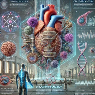
The principle of structure-function relationship is central to biology and physiology, revealing how the physical configuration of a biological component directly influences its performance. From the molecular level to entire organ systems, the way something is built often dictates how it works. To truly appreciate this connection, learners begin with foundational concepts in science, particularly the study of biology, which provides an integrated understanding of life’s complexity.
Understanding cell biology is essential for exploring how the structural attributes of cells, such as membrane shape or organelle arrangement, influence processes like transport, communication, and energy transformation. Key topics such as cell structure and cell physiology show how components like mitochondria and ribosomes are specifically designed for their metabolic or synthetic roles.
During development, cells undergo remarkable changes in form, a process explored in cell development. These changes support new functions and reflect the adaptability inherent in living systems. Likewise, the cell cycle illustrates how nuclear and cytoskeletal structures coordinate to ensure accurate cell division.
On the intercellular scale, cell communication relies on receptor and signal molecule structures that fit together with precision, akin to a lock-and-key mechanism. This understanding supports exploration of higher-order signaling in signal communication and the maintenance of homeostasis, both of which depend on finely tuned physiological mechanisms.
The broader scope of physiology highlights examples like the alveoli of the lungs, whose thin, moist walls are ideally suited for gas exchange, and the structure of skeletal muscle fibers that enables rapid contraction. Adaptations that reinforce this principle are explored in adaptation and plasticity, where organisms evolve structural modifications to meet environmental challenges.
Metabolic processes, described in energy and metabolism, further demonstrate the importance of enzyme structure in catalyzing specific biochemical reactions. These enzymes function based on their active site architecture, which binds substrates in highly selective ways.
At the molecular scale, the arrangement of nucleotides in DNA and RNA facilitates accurate replication and transcription, supporting functions from protein synthesis to gene expression. Any alterations in structure, such as in genetic mutation, can drastically impact function and potentially lead to disease.
This functional sensitivity to structure is a recurring theme in molecular genetics. Concepts such as DNA technology and applications of genetics in medicines exploit the structural nature of biological molecules to develop diagnostics and treatments.
Even across evolutionary timescales, structural changes are at the heart of molecular evolution. The variation in form across populations links to functional outcomes studied in genetics, molecular genetics, and broader ecological contexts through ecology and evolutionary biology.
The structure-function principle ties together many biological disciplines, from molecular biology to systems physiology. It remains a guiding framework in gene expression, molecular genetics, and applications in medicine, illustrating the elegance and interdependence of biological design and function.
IDEF0 Control Architecture: Structure-Function Optimization Framework
INPUT: Raw Biomolecular Substrates, Mechanical Forces, Environmental Energy Inputs & Hydrodynamic Pressures
CONTROL: Genomic Code Constraints, Thermodynamic Stability Rules, Evolutionary Selection Pressures & Allosteric Boundaries
MECHANISM: 3D Protein Folding Configurations, Cytoskeletal Networks, Specialized Tissue Histology & Anatomical Biomechanics
OUTPUT: Specialized Biological Function (Substrate Catalysis, Force Generation, Gas Exchange, Rapid Signal Conduction)
Physiology – Overview
Frames biological function as emerging from structural organisation across cells, tissues, and organ systems. Explore Overview →
Structure–Function Relationship (Current Page)
Examines how anatomical features at all scales enable specific physiological roles and system-level performance.
Homeostasis
Depends on specialised structures that support efficient regulation and feedback control mechanisms. Learn Homeostasis →
Energy Metabolism
Relies on cellular and organ structures optimised for energy production and utilisation. Explore Energy & Metabolism →
Signal Communication
Uses specialised structural features such as receptors and neural networks to transmit information effectively. Discover Signal Communication →
Adaptation & Plasticity
Demonstrates how structural changes over time enable functional adaptation to environmental demands. Study Adaptation & Plasticity →
Molecular Level: Structure Determines Function
At the molecular level, the three-dimensional structure of biological molecules, such as proteins and nucleic acids, determines their functions:
Enzymes and Substrates
- Enzymes have a specific active site where substrates bind, facilitating biochemical reactions.
- The lock-and-key model or the induced fit model describes how the structure of the enzyme ensures specificity.
- Example: Hexokinase, a key enzyme in glycolysis, has a structure that allows it to bind glucose and phosphorylate it efficiently.
DNA and Its Double Helix
- The double-helical structure of DNA allows for the storage, replication, and transmission of genetic information.
- Complementary base pairing (A-T and G-C) ensures accurate replication during cell division.
Hemoglobin
- Hemoglobin’s quaternary structure (composed of four polypeptide chains) enables it to bind oxygen efficiently.
- Structural changes upon oxygen binding (cooperative binding) optimize oxygen uptake and delivery to tissues.
Cellular Level: Form and Function in Cells
At the cellular level, the shapes and internal structures of cells are specialized to fulfill specific functions:
Red Blood Cells (Erythrocytes)
- Red blood cells have a biconcave shape that increases surface area for oxygen diffusion and allows flexibility to pass through narrow capillaries.
- The absence of a nucleus provides more space for hemoglobin, maximizing oxygen transport.
Neurons
- Neurons have long, thin axons for transmitting electrical signals over long distances and dendrites for receiving inputs from other neurons.
- This structure enables rapid and efficient communication within the nervous system.
Muscle Cells
- Muscle cells (fibers) are elongated and contain parallel myofibrils composed of actin and myosin filaments.
- The arrangement of these contractile proteins allows for coordinated contraction and force generation.
Intestinal Epithelial Cells
- These cells have specialized microvilli on their surface to increase surface area for nutrient absorption.
Tissue Level: Specialization for Function
At the tissue level, the organization of cells into functional units optimizes physiological processes:
Epithelial Tissue
- The tightly packed cells of epithelial tissue form protective barriers and regulate exchange.
- Example: The alveolar epithelium in the lungs is thin to facilitate rapid gas exchange.
Connective Tissue
- Connective tissues like bone and cartilage provide structural support.
- Example: The honeycomb structure of spongy bone reduces weight while maintaining strength for movement and protection.
Muscle Tissue
- Striated muscle tissue (skeletal muscle) is organized in fibers with aligned actin and myosin filaments to generate force.
- Cardiac muscle has intercalated discs to synchronize heart contractions.
Nervous Tissue
- Nervous tissue consists of neurons and glial cells, organized to transmit and process information.
Organ Level: Structural Adaptations for Function
At the organ level, structures are designed to perform complex physiological functions:
Lungs
- The lungs have a highly branched structure that culminates in millions of alveoli.
- This design maximizes surface area for efficient gas exchange.
Heart
- The heart’s muscular walls and specialized chambers are designed for pumping blood.
- Valves prevent backflow, ensuring unidirectional blood flow.
Kidneys
- The kidney’s nephrons (functional units) filter blood, regulate ion balance, and remove waste.
- The loop of Henle is elongated in desert animals to conserve water, demonstrating an adaptation to their environment.
Small Intestine
- The inner lining of the small intestine contains villi and microvilli to maximize nutrient absorption.
Brain
- The brain’s folded surface (gyri and sulci) increases surface area, allowing for greater processing power.
System Level: Integration of Structure and Function
At the system level, multiple organs work together to achieve higher-order functions:
Circulatory System
- The heart pumps blood through a network of arteries, veins, and capillaries.
- Capillaries are thin-walled to allow nutrient and gas exchange at the tissue level.
Respiratory System
- The branching airways and alveolar sacs are designed to maximize oxygen uptake and carbon dioxide removal.
Musculoskeletal System
- Bones provide a rigid structure for support and movement, while muscles attach to bones through tendons for force generation.
Nervous System
- The central and peripheral nervous systems enable communication between the brain and body, facilitating rapid responses.
Structural Adaptations in Animals and Plants
The relationship between structure and function is also evident in adaptations to specific environments:
Animals
- Birds have hollow bones to reduce weight for flight.
- Whales have streamlined bodies and thick blubber for buoyancy and insulation in water.
- Cheetahs have elongated limb bones and flexible spines for running at high speeds.
Plants
- Leaves have large surface areas for maximum light absorption during photosynthesis.
- The stomata allow gas exchange but can close to prevent water loss.
- Xylem vessels are long, hollow tubes that facilitate water transport from roots to leaves.
Clinical and Biomedical Relevance of Structure-Function Relationship
Understanding the structure-function relationship is critical for medicine and biomedical research:
- Diseases: Structural abnormalities often underlie disease processes.
- Medical Devices: Devices like heart valves and prosthetic limbs are designed to mimic the structure-function relationship of biological tissues.
- Drug Design: Understanding protein structures enables the development of drugs that target specific sites (e.g., enzymes, receptors).
Why Study Structure-Function Relationship
Core Principle of Biology
Structure-function relationship explains how the shape and arrangement of biological components determine their roles. Students learn that structure is not arbitrary but closely linked to function. This principle applies to molecules, cells, organs, and systems. It is central to understanding biological design and evolution.
Examples Across Biological Levels
From the double helix of DNA to the folding of proteins and the shape of red blood cells, structure supports function. Students study how anatomy, histology, and biochemistry reveal form-function correlations. These examples help link theory to observation. They enhance appreciation of life’s complexity.
Implications in Health and Disease
Altered structures can lead to malfunction and disease, such as misfolded proteins in Alzheimer’s. Understanding structure-function links helps explain symptoms and guide treatments. It also supports innovations in drug design and biomaterials. Students see the importance of this relationship in real-world contexts.
Design Principles in Engineering and Technology
Biological structure-function insights inspire innovations in biomimicry and biotechnology. Students apply these concepts in designing medical devices and synthetic systems. This bridges biology and engineering disciplines. It encourages creative thinking and innovation.
Support for Research and Diagnostics
Analyzing how structure affects function is key to many experimental approaches. Students use microscopy, modeling, and imaging tools to investigate form and function. These skills are essential in diagnostics and research. They form the basis of a well-rounded scientific education.
About This Interactive Simulator:
This simulator models the enzyme-substrate catalytic rate based on active site structural binding affinity and surface-area-to-volume transport kinetics. Adjust the binding geometry (induced fit) and membrane surface area parameters to evaluate reaction velocity and diffusion flux in real time.
- Active Site Complementarity Slider: Modifies active site structural alignment to observe shifts in substrate binding affinity (Km) and catalytic turnover rate.
- Surface Area Optimization Slider: Simulates structural folding (villi/microvilli) to demonstrate how surface-area-to-volume ratio scales biological flux.
- Real-Time Reaction Kinetics Canvas: Displays substrate concentration versus reaction velocity curves (Michaelis-Menten dynamics) with axis tick labels and maximum velocity (Vmax) thresholds.
Interactive Enzyme Kinetics & Structural Optimization Simulator
Applications of Structure-Function Relationship in Biomedicine and Engineering
The structure-function principle forms the foundational pillar of modern rational drug design and tissue engineering. By determining the 3D atomic structure of biological targets using X-ray crystallography and cryo-EM, pharmacologists synthesize small molecules that fit active sites with high precision.
In biomedical engineering, artificial heart valves are constructed with tri-leaflet geometry to replicate natural hemodynamics, preventing turbulent blood flow and reducing clot formation. Similarly, synthetic vascular grafts feature microporous surface architectures that encourage endothelial cell attachment and integration.
In genetic medicine, understanding how misfolding mutations alter tertiary protein structure allows developers to design chemical chaperones. These therapeutic molecules stabilize mutant proteins, restoring normal cellular transport and functional activity.
Emerging Trends, Challenges & Future Directions
Advances in artificial intelligence and deep learning models (such as AlphaFold) have revolutionized structural biology by predicting 3D protein structures directly from primary amino acid sequences. This capability accelerates target identification and bio-molecular design.
A persistent challenge remains modeling dynamic structural transitions in real time. Static crystal structures do not fully capture the conformational flexibility and allosteric movements that dictate biological function in living systems.
Future directions lie in de novo protein design, where synthetic proteins with novel active sites and structural motifs are engineered from scratch to perform custom catalytic reactions, synthesize biofuels, or neutralize environmental toxins.
Frequently Asked Questions
What does structure–function relationship mean in physiology?
In physiology, the structure–function relationship describes how the shape, organisation, and material properties of cells, tissues, and organs are directly linked to what they do. The way something is built — from membranes and proteins to whole organ systems — helps explain how it performs its physiological role.
Why is the structure–function relationship important for understanding the human body?
The structure–function relationship is important because it connects anatomy to physiology. By understanding how tissues and organs are arranged, what they are made of, and how they are shaped, we can better explain how they generate force, transport substances, transmit signals, or regulate body conditions in real time.
Can you give simple examples of structure–function relationships in physiology?
Examples include the thin, moist alveoli in the lungs that maximise surface area for gas exchange; the biconcave shape of red blood cells that increases surface area and flexibility for squeezing through capillaries; and the long, insulated axons of neurons that allow rapid electrical signalling over long distances.
How does structure–function apply at the cellular and molecular levels?
At the cellular and molecular levels, structure–function appears in the folding of proteins that creates specific active sites, the arrangement of ion channels in membranes that controls electrical activity, and the organisation of cytoskeleton fibres that support cell shape and movement. Small changes in structure can greatly alter function.
What happens when structure is altered by injury or disease?
When structure is altered by injury, genetic mutation, or disease, function is often impaired. For example, scarring of lung tissue can reduce elasticity and gas exchange, damaged heart valves can disrupt blood flow, and misfolded proteins can lead to loss of enzyme activity or toxic aggregations in nerve cells.
How do engineers and clinicians use structure–function principles?
Engineers and clinicians use structure–function principles to design medical devices, prosthetic joints, stents, and artificial valves that mimic or support natural function. Clinicians also interpret imaging and test results by linking structural changes on scans or histology slides to likely functional consequences for the patient.
How does the structure–function relationship help explain adaptation and performance?
Structure–function relationships help explain how training, growth, or evolution improves performance. For instance, endurance training can increase capillary density and mitochondrial content in muscles, while evolutionary changes in heart size or limb structure can support faster running, deeper diving, or more efficient flight in different species.
Why is understanding structure–function relationships useful for future studies and careers?
Understanding structure–function relationships prepares students for university studies in medicine, biomedical science, physiotherapy, sports science, and bioengineering. It is also valuable in careers that involve interpreting scans, designing medical technologies, analysing movement, or explaining how lifestyle and disease alter the body’s structure and performance.
End-of-Page Practice & Assessment Module
Section 1: Foundational Concept Review Questions
-
Question 1: How does the active site geometry of an enzyme dictate its catalytic specificity?
Answer: An enzyme's active site possesses a unique 3D arrangement of amino acid residues that match the shape, charge distribution, and hydrophobicity of its substrate. This complementary structural fit lowers activation energy by stabilizing the transition state during catalysis. -
Question 2: How does the biconcave architecture of an erythrocyte enhance respiratory gas exchange?
Answer: The biconcave disk shape increases the cell's surface-area-to-volume ratio compared to a sphere of equal volume, shortening the diffusion distance for oxygen and CO2. It also provides membrane flexibility for navigating capillary beds. -
Question 3: How does the quaternary structure of hemoglobin enable cooperative oxygen binding?
Answer: Hemoglobin consists of four polypeptide subunits (α2β2). Binding of oxygen to one heme group induces a conformational shift from the T (tense) state to the R (relaxed) state, increasing affinity for oxygen at the remaining binding sites. -
Question 4: What structural features of the alveolar-capillary membrane optimize gas diffusion rate?
Answer: The membrane consists of simple squamous Type I pneumocytes fused to capillary endothelial cells, creating an ultra-thin barrier (≈ 0.5 μm) across a cumulative surface area of nearly 100 m2 in adult lungs. -
Question 5: How does the structural organization of sarcomeres generate mechanical force in skeletal muscle?
Answer: Sarcomeres contain overlapping thick (myosin) and thin (actin) myofilaments anchored between Z-discs. Cross-bridge cycling pulls actin filaments toward the M-line, shortening the sarcomere and producing linear force. -
Question 6: How does the structural length of the Loop of Henle correlate with renal concentrating ability?
Answer: Longer loops of Henle extend deeper into the hyperosmotic renal medulla, creating a steeper hypertonic gradient that drives greater water reabsorption from collecting ducts, conserving body water in arid environments. -
Question 7: How do intercalated discs structurally coordinate cardiac muscle contraction?
Answer: Intercalated discs contain desmosomes for mechanical anchoring during force generation and gap junctions for low-resistance electrical coupling, allowing action potentials to spread rapidly for synchronized atrial and ventricular contraction. -
Question 8: How does the structural arrangement of villi and microvilli support intestinal nutrient absorption?
Answer: Folding of the mucosa into villi and brush border microvilli expands the intestinal surface area up to 600-fold, maximizing space for digestive enzymes and nutrient transport proteins.
Section 2: Analytical & Scenario-Based Thought Questions
-
Question 1: How does a single amino acid substitution alter hemoglobin structure and result in sickle cell anemia?
Answer: Substituting hydrophilic glutamic acid with hydrophobic valine at position 6 of the β-globin chain creates a hydrophobic patch. Under deoxygenated conditions, these patches aggregate into long rigid polymers that distort red blood cells into a sickle shape, causing vascular occlusion and hemolysis. -
Question 2: Why does pulmonary fibrosis impair gas exchange even if total lung surface area remains unchanged?
Answer: Fibrosis causes collagen deposition that thickens the alveolar-capillary basement membrane. According to Fick's Law of Diffusion, increasing diffusion distance directly reduces diffusion rate, leading to hypoxemia. -
Question 3: How does the double-helix geometry of DNA protect genetic information while enabling replication?
Answer: Nitrogenous bases are tucked inside the hydrophobic core, protected from chemical damage by the exterior sugar-phosphate backbone. Complementary base pairing provides redundant sequence information on opposite strands for semi-conservative replication. -
Question 4: How does myocyte hypertrophy alter cardiac structure and functional stroke volume during chronic hypertension?
Answer: Pumping against elevated systemic vascular resistance causes concentric left ventricular hypertrophy. Thicker muscular walls maintain stroke volume initially, but reduced ventricular lumen volume and compliance eventually restrict diastolic filling. -
Question 5: How do myelin sheath structural disruptions in Multiple Sclerosis affect action potential propagation?
Answer: Demyelination removes high electrical resistance insulation across internodes, allowing ionic current to leak. Current density at subsequent Nodes of Ranvier fails to reach threshold, causing conduction block or slowing signal transmission. -
Question 6: How does the structural folding (gyri and sulci) of the human cerebral cortex expand cognitive capacity?
Answer: Gyri and sulci allow a larger surface area of gray matter (neuronal cell bodies) to fit within the fixed volume of the cranium, expanding cortical processing density without requiring a disproportionately large skull. -
Question 7: How do competitive and non-competitive enzyme inhibitors alter active site structure and reaction kinetics differently?
Answer: Competitive inhibitors structurally resemble the substrate and bind directly to the active site without changing Vmax. Non-competitive inhibitors bind to allosteric sites, altering the enzyme's 3D conformation and reducing Vmax regardless of substrate concentration. -
Question 8: How do plant xylem vessels structurally resist collapse during high transpirational pull?
Answer: Xylem vessel elements are reinforced with secondary cell walls containing rigid lignin polymers arranged in rings or helices. This structural reinforcement withstands extreme negative hydrostatic pressures (tension) without collapsing.
Section 3: Numerical & Quantitative Physiology Calculations
-
Problem 1: Calculate the total surface area in m2 available for gas exchange in a lung containing 300 million alveoli, assuming each alveolus is a sphere with an average radius r = 100 μm. (Formula: Surface Area of sphere = 4 × π × r2).
Solution:
r = 100 μm = 100 × 10-6 m = 10-4 m.
Surface area of 1 alveolus = 4 × π × (10-4 m)2 = 4 × 3.14159 × 10-8 m2 ≈ 1.2566 × 10⁻⁷ m2.
Total Surface Area = 300,000,000 × 1.2566 × 10-7 m2 = 37.70 m2.
The total alveolar gas exchange surface area is approximately 37.70 m2. -
Problem 2: According to Fick's Law of Diffusion, Gas Diffusion Rate J = (A × D × ΔP) / Δx. Calculate the percentage change in oxygen diffusion rate if pulmonary edema increases the membrane thickness (Δx) from 0.5 μm to 2.0 μm, assuming area (A), diffusion constant (D), and pressure gradient (ΔP) remain constant.
Solution:
Initial diffusion rate J1 ∝ 1 / 0.5 = 2.0.
New diffusion rate J2 ∝ 1 / 2.0 = 0.5.
Percentage change = ((0.5 - 2.0) / 2.0) × 100 = (-1.5 / 2.0) × 100 = -75%.
Oxygen diffusion rate decreases by 75% due to increased membrane thickness. -
Problem 3: Calculate the surface-area-to-volume ratio (SA/V in μm-1) for a spherical cell of radius r = 10 μm compared to a biconcave erythrocyte model simplified as a flattened cylinder of radius r = 4 μm and height h = 1 μm. (Spherical SA = 4πr2, V = (4/3)πr3; Cylinder SA = 2πr2 + 2πrh, V = πr2h).
Solution:
Spherical Cell: SA/V = (4πr2) / ((4/3)πr3) = 3 / r = 3 / 10 = 0.30 μm-1.
Flattened Cell: SA = 2π(4)2 + 2π(4)(1) = 32π + 8π = 40π ≈ 125.66 μm2.
V = π(4)2(1) = 16π ≈ 50.27 μm3.
SA/V = 40π / 16π = 2.50 μm-1.
The flattened model yields a surface-area-to-volume ratio of 2.50 μm-1 versus 0.30 μm-1 for the sphere, demonstrating an 8.33-fold structural optimization. -
Problem 4: Using Poiseuille's Law, Resistance R = (8 × η × L) / (π × r4), calculate the fold-change in vascular resistance if arteriole vasoconstriction reduces its internal vessel radius (r) by 20% (new radius r' = 0.8 × r).
Solution:
Rinitial ∝ 1 / r4.
Rnew ∝ 1 / (0.8 × r)4 = 1 / (0.4096 × r4) = 2.4414 × Rinitial.
A 20% reduction in vessel radius increases vascular resistance by approximately 2.44-fold (144.14% increase). -
Problem 5: In an enzyme kinetics experiment following Michaelis-Menten dynamics v = (Vmax × [S]) / (Km + [S]), calculate the reaction velocity v in μmol/min when substrate concentration [S] = 12 mM, given Vmax = 100 μmol/min and Km = 4 mM.
Solution:
v = (100 × 12) / (4 + 12)
v = 1200 / 16 = 75 μmol/min.
The reaction velocity is 75 μmol/min at 12 mM substrate concentration. -
Problem 6: Calculate the fold expansion in mucosal surface area achieved by intestinal villi and microvilli if primary circular folds increase surface area 3-fold, villi increase it by an additional 10-fold, and microvilli increase it by a further 20-fold.
Solution:
Total Fold Expansion = 3 × 10 × 20 = 600-fold.
The structural mucosal modifications expand the total absorptive surface area 600-fold. -
Problem 7: A skeletal muscle fiber has an initial resting sarcomere length of 2.5 μm. During maximal contraction, each sarcomere shortens to 1.8 μm. Calculate the total percentage shortening of a 10 cm long muscle fiber assuming uniform sarcomere contraction along its length.
Solution:
Sarcomere shortening = 2.5 μm - 1.8 μm = 0.7 μm.
Percentage shortening = (0.7 μm / 2.5 μm) × 100 = 28%.
Because contraction is uniform across all series sarcomeres, the 10 cm muscle fiber shortens by 28% to a final length of 7.2 cm.